. 2024 Aug 1;4(8):1894-1907.
doi: 10.1158/2767-9764.CRC-24-0124.

p300 KAT Regulates SOX10 Stability and Function in Human Melanoma

Affiliations

p300 KAT Regulates SOX10 Stability and Function in Human Melanoma

Aaron Waddell et al. Cancer Res Commun. .

Abstract

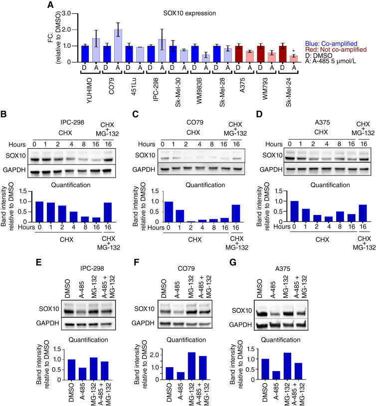
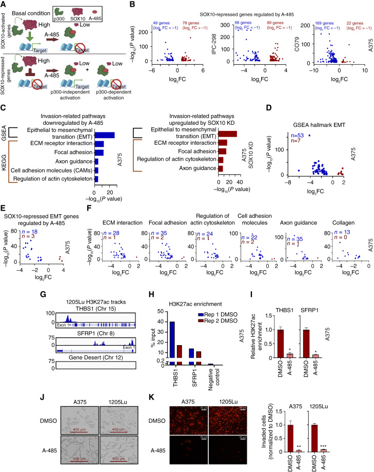
SOX10 is a lineage-specific transcription factor critical for melanoma tumor growth; on the other hand, SOX10 loss-of-function drives the emergence of therapy-resistant, invasive melanoma phenotypes. A major challenge has been developing therapeutic strategies targeting SOX10's role in melanoma proliferation while preventing a concomitant increase in tumor cell invasion. In this study, we report that the lysine acetyltransferase (KAT) EP300 and SOX10 gene loci on chromosome 22 are frequently co-amplified in melanomas, including UV-associated and acral tumors. We further show that p300 KAT activity mediates SOX10 protein stability and that the p300 inhibitor A-485 downregulates SOX10 protein levels in melanoma cells via proteasome-mediated degradation. Additionally, A-485 potently inhibits proliferation of SOX10+ melanoma cells while decreasing invasion in AXLhigh/MITFlow melanoma cells through downregulation of metastasis-related genes. We conclude that the SOX10/p300 axis is critical to melanoma growth and invasion and that inhibition of p300 KAT activity through A-485 may be a worthwhile therapeutic approach for SOX10-reliant tumors.

Significance: The p300 KAT inhibitor A-485 blocks SOX10-dependent proliferation and SOX10-independent invasion in hard-to-treat melanoma cells.


Conflict of interest statement

P.A. Cole is a cofounder of Acylin Therapeutics and reports personal fees from Acylin Therapeutics and AbbVie during the conduct of the study; personal fees from Scorpion Therapeutics and Nested Therapeutics outside the submitted work; and a patent for Methods and Compositions for Modulating P300/CBP Activity issued, licensed, and with royalties paid. R.M. Alani is a cofounder of Acylin Therapeutics and reports personal fees from Acylin Therapeutics prior to the conduct of this study. Her spouse, P.A. Cole, has disclosures as noted above. No disclosures were reported by the other authors.

Figures

Figure 1
Figure 1
EP300 is commonly co-amplified with SOX10 in melanomas. A, The EP300 and SOX10 genes are located in close proximity on chromosome 22. B,EP300 and SOX10 amplification prevalence in different melanoma datasets [TCGA, Dana Farber Cancer Institute (DFCI), Memorial Sloan Kettering Cancer Center (MSKCC)]. Acral patients from multiple datasets were combined (combined acral). Amplification levels were defined using GISTIC2.0. C,EP300 (left) and SOX10 (right) expressions were analyzed for each copy-number level, as defined by GISTIC2.0 (data from TCGA; Del, deletion; D, diploid; low, low-level amplification; high, high-level amplification). Statistical analysis is compared with diploid samples. D,EP300 and SOX10 copy numbers are positively correlated in melanoma cell lines (data from the CDM). E,EP300 copy number and expression are positively correlated in melanoma cell lines (left). SOX10 copy number and expression are positively correlated in melanoma cell lines (right; data CDM). F,EP300/SOX10 co-amplifications (>2 copies of EP300 and SOX10) are common in melanoma cell lines (data from CDM). *, P < 0.05; **, P < 0.005; ***, P < 0.0005.
Figure 2
Figure 2
SOX10 signaling requires p300 KAT activity. (A) A panel of melanoma cell lines were treated with DMSO (D) or 5 µmol/L A-485 (A) for 24 hours. SOX10, MITF, and DCT protein levels were assessed via immunoblotting. B, A panel of EP300/SOX10 co-amplified melanoma cell lines were treated with DMSO (D) or 5 µmol/L A-485 (A) for 24 hours. MITF and DCT expressions were assessed via RT-qPCR. C,MITF, SCD, and MYC expressions were downregulated by SOX10 KD in A375 cells. D, A panel of melanoma cell lines without EP300/SOX10 co-amplifications were treated with DMSO (D) or 5 µmol/L A-485 (A) for 24 hours. MITF, SCD, and MYC expressions were assessed via RT-qPCR. *, P < 0.05; **, P < 0.005; ***, P < 0.0005.
Figure 3
Figure 3
A-485 induces proteasomal degradation of SOX10. A, A panel of melanoma cell lines were treated with DMSO (D) or 5 µmol/L A-485 (A) for 24 hours. SOX10 expression was assessed via RT-qPCR. B–D, IPC-298, CO79, and A375 cells were treated with 50 µg/mL CHX and/or 5 µmol/L MG-132 for the indicated time. SOX10 protein levels were assessed via immunoblotting. E–G, IPC-298, CO79, and A375 cells were treated with DMSO, 5 µmol/L A-485, 5 µmol/L MG-132, or their combination for 16 hours. SOX10 protein levels were assessed via immunoblotting. *, P < 0.05.
Figure 4
Figure 4
A-485 treatment potently downregulates SOX10 target genes in melanoma cells. A–C, IPC-298, CO79, and A375 cells were treated with 5 µmol/L A-485 for 24 hours, and RNA-seq was performed. Volcano plots of DEGs in each cell line are depicted (DEGs defined as FC > |2| and P < 0.05). D, The top five GO biological process pathways are shown for downregulated genes in each cell line. E, A heatmap of DEGs resulting from SOX10 KD in A375 cells is shown (data from GSE50535). F, The top five GO biological process pathways are shown for SOX10-activated genes (i.e., genes downregulated by SOX10 KD). G, A-485 downregulates GO biological process pathways that are activated by SOX10 in IPC-298, CO79, and A375 cells. H, Volcano plots of SOX10-activated genes differentially expressed due to A-485 treatment for IPC-298, CO79, and A375 cells. I–K, GO biological process pathways are shown for SOX10-activated genes that are downregulated by A-485.
Figure 5
Figure 5
A-485 potently inhibits proliferation of SOX10+ melanoma cell lines. A, A panel of melanoma cell lines were treated with A-485 (in a range of doses) for 6 days, and their relative proliferation vs. control was assessed (left). The results for 5 µmol/L A-485 vs. control are shown (right). B, Cells that did not respond to A-485 within 6 days were pretreated with 5 µmol/L A-485 for 9 days and reseeded for a 3-day proliferation assay, and their relative proliferation vs. control was assessed (total 12 days of drug treatment). C, WM983B-R and SK-Mel-28-R BRAFi-resistant cells have low expression of SOX10 in comparison with parental cells. D, The proliferation effects of short-term (6-day) or long-term (12-day) A-485 treatment of WM983B-R and SK-Mel-28-R cells were assessed. E, 451Lu-R cells maintain SOX10 expression vs. BRAFi-sensitive parental cells (left). 451Lu-S and 451Lu-R were treated with A-485 (in a range of doses) for 6 days, and their relative proliferation vs. control was assessed (right). F, A375-R cells maintain SOX10 expression vs. BRAFi-sensitive parental cells (left). A375-S and A375-R cells were treated with A-485 (in a range of doses) for 6 days, and their relative proliferation vs. control was assessed (middle). A375-S and A375-R cells were pretreated 5 µmol/L A-485 for 9 days and reseeded for a 3-day proliferation assay, and their relative proliferation vs. control was assessed (right). G, EP300 dependency scores are plotted for melanoma cell lines with low SOX10 [<4 log2(TPM + 1)] vs. high SOX10 [>4 log2(TPM + 1)] expression in the CDM 22Q2 public + score, Chronos dataset. *, P < 0.05; **, P < 0.005; ***, P < 0.0005.
Figure 6
Figure 6
p300 KAT activity is essential for the activation of SOX10-repressed EMT markers. A, Diagram depicting the hypothesis that p300 activity is essential for the expression of SOX10-activated genes, but SOX10-repressed genes may also require p300 for their activation. B, Volcano plots of SOX10-repressed genes differentially expressed due to A-485 treatment for IPC-298, CO79, and A375 cells (DEGs defined as FC > |2| and P < 0.05). C, SOX10-repressed genes are enriched for genes involved in the EMT, and similar invasion-related pathways are downregulated by A-485. CAM, cell adhesion molecules; ECM, extracellular matrix. D, Volcano plot of EMT genes differentially expressed due to A-485 treatment for A375 cells. E, Volcano plot of SOX10-regulated EMT genes differentially expressed due to A-485 treatment for A375 cells. F, Volcano plots of genes differentially expressed due to A-485 are shown for various KEGG invasion-related pathways identified in C. Differentially expressed collagen genes are also shown. G, H3K27ac is enriched at the promoters of EMT genes THBS1 and SFRP1 in comparison with a gene desert in invasive 1205Lu cells via ChIP sequencing. H, H3K27ac was confirmed to be enriched at the promoters of THBS1 and SFRP1 in comparison with a gene desert in A375 cells via ChIP-qPCR. I, A-485 decreases H3K27ac enrichment at the promoters of THBS1 and SFRP1, as assessed by ChIP-qPCR. (J) 5 µmol/L A-485 alters the cell morphology of A375 and 1205Lu cells after long-term (12-day) treatment. K, A-485 potently inhibits invasion in A375 and 1205Lu after long-term (12-day) treatment. Representative images of invaded cells are shown on the left, and quantification of invaded cells is shown on the right. *, P < 0.05; **, P < 0.005; ***, P < 0.0005.

Update of

Similar articles

Cited by

References

    1. Guo W, Wang H, Li C. Signal pathways of melanoma and targeted therapy. Signal Transduct Target Ther 2021;6:424. - PMC - PubMed
    1. Tang Y, Durand S, Dalle S, Caramel J. EMT-inducing transcription factors, drivers of melanoma phenotype switching, and resistance to treatment. Cancers (Basel) 2020;12:2154. - PMC - PubMed
    1. Najem A, Soumoy L, Sabbah M, Krayem M, Awada A, Journe F, et al. . Understanding molecular mechanisms of phenotype switching and crosstalk with TME to reveal new vulnerabilities of melanoma. Cells 2022;11:1157. - PMC - PubMed
    1. Tsoi J, Robert L, Paraiso K, Galvan C, Sheu KM, Lay J, et al. . Multi-stage differentiation defines melanoma subtypes with differential vulnerability to drug-induced iron-dependent oxidative stress. Cancer Cell 2018;33:890–904.e5. - PMC - PubMed
    1. Shakhova O, Zingg D, Schaefer SM, Hari L, Civenni G, Blunschi J, et al. . Sox10 promotes the formation and maintenance of giant congenital naevi and melanoma. Nat Cell Biol 2012;14:882–90. - PubMed

MeSH terms