. 2024 Sep;11(35):e2402284.
doi: 10.1002/advs.202402284. Epub 2024 Jul 12.

Hyperactivation of mTOR/eIF4E Signaling Pathway Promotes the Production of Tryptophan-To-Phenylalanine Substitutants in EBV-Positive Gastric Cancer

Affiliations

Hyperactivation of mTOR/eIF4E Signaling Pathway Promotes the Production of Tryptophan-To-Phenylalanine Substitutants in EBV-Positive Gastric Cancer

Zi-Qi Zheng et al. Adv Sci (Weinh). 2024 Sep.

Abstract

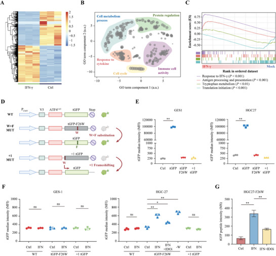
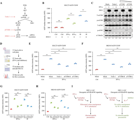
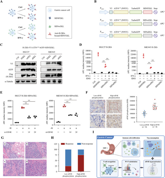
Although messenger RNA translation is tightly regulated to preserve protein synthesis and cellular homeostasis, chronic exposure to interferon-γ (IFN-γ) in several cancers can lead to tryptophan (Trp) shortage via the indoleamine-2,3-dioxygenase (IDO)- kynurenine pathway and therefore promotes the production of aberrant peptides by ribosomal frameshifting and tryptophan-to-phenylalanine (W>F) codon reassignment events (substitutants) specifically at Trp codons. However, the effect of Trp depletion on the generation of aberrant peptides by ribosomal mistranslation in gastric cancer (GC) is still obscure. Here, it is shows that the abundant infiltrating lymphocytes in EBV-positive GC continuously secreted IFN-γ, upregulated IDO1 expression, leading to Trp shortage and the induction of W>F substitutants. Intriguingly, the production of W>F substitutants in EBV-positive GC is linked to antigen presentation and the activation of the mTOR/eIF4E signaling pathway. Inhibiting either the mTOR/eIF4E pathway or EIF4E expression counteracted the production and antigen presentation of W>F substitutants. Thus, the mTOR/eIF4E pathway exposed the vulnerability of gastric cancer by accelerating the production of aberrant peptides and boosting immune activation through W>F substitutant events. This work proposes that EBV-positive GC patients with mTOR/eIF4E hyperactivation may benefit from anti-tumor immunotherapy.

Keywords: EBV‐positive gastric cancer; aberrant mRNA translation; codon reassignment; substitutants; tryptophan.


Conflict of interest statement

The authors declare no conflict of interest.

Figures

Figure 1
Figure 1
IFN‐γ upregulated IDO1 expression and caused tryptophan exhaustion in EBV‐positive gastric cancer. A) The model diagram showing the effect of IFN‐γ on the generation of ribosomal abnormal peptides. B) The representative images (up panel) and quantification results (bottom panel) of multiplex immunofluorescence staining showing the CD20+ B cell, CD4+ and CD8+ T cell infiltration in 8 pairs of EBV‐negative (NT1‐8) and EBV‐positive (PT1‐8) GC tissues. C) Correlation analysis between mRNA expression of IFNG and IDO1 in TCGA cohort of stomach adenocarcinoma (STAD) patients (n = 375). D) Representative images showing IDO1 staining in EBV‐positive GC tissues (n = 8, PT1‐8) and paired EBV‐negative GC tissues (n = 8, NT1‐8) (left panel). The integrated optical density (IOD) of positive expression cells in 10 cases of GC tissues, and grading of the level of immunoreactivity (right panel). Scale bar = 100 µm. E) The expression level of IDO1 protein was quantified by Western blotting in 5 pairs of EBV‐positive (PT1‐5) and EBV‐negative GC tissues (NT1‐5). F) The expression of IDO1 in other 6 pairs of EBV‐positive and EBV‐negative GC tissues as determined by qPCR. G) Comparison of IDO1 expression level between EBV‐negative GC (n = 74) and EBV‐negative GC (n = 6) using the gene expression profile data from GSE122401 (n = 80). H) Western blotting evaluation of the changes in IDO1 expression in HGC27, MKN45, AGS and GES1 cells in response to IFN‐γ treatment. I, J) The tryptophan (I) and kynurenine (J) levels in HGC27 and MKN45 cells under mock or IFN‐γ treatment were determined using LC‐MS/MS. Data are presented as the mean ± SD. *P < 0.05, **P < 0.01.
Figure 2
Figure 2
Tryptophan deficiency resulted in tryptophan‐to‐phenylalanine substitution translation in GC cells. A) Cluster heatmap of the differentially expressed genes in IFN‐γ treated MKN45 cells compared to the mock treated MKN45 cells. B) REVIGO[ 48 ] summary of the different Gene Ontology (GO) terms between mock‐ and IFN‐γ treated MKN45 cells. C) GSEA plots of the enrichment pathway associated with IFN‐γ treatment in MKN45 cells. D) This figure illustrates the constructs utilized to identify frameshifting and substitution translation events in GC cell. The Wild Type (WT) vector, containing a V5‐tag (blue box), was linked to ATF41–63 (red box, featuring tryptophan residue W at the 93rd amino acid from the translation starting site) and positioned upstream of TurboGFP (tGFP, green box, with fewer tryptophan residues). The W>F substitution construct (W>F MUT) was derived from the WT vector, where the phenylalanine residue at position 26 was altered to tryptophan (F26W). The constructs for detecting frameshifting events (+1 MUT) were also transformed from the WT, with one additional base pair upstream the tGFP and leading to an out‐of‐frame tGFP product. E) The tGFP signals in GES1 and HGC27 cells stably expressing WT, W>F MUT, or +1 MUT reporters, respectively, were measured by flow cytometry assay. F, G) GES1 and HGC27 cells stably expressing the WT, W>F MUT, or +1 MUT reporters were mock‐treated or treated with IFN‐γ (250U/mL) for 48 h and subjected for flow cytometry analysis (F) or mass spectrometry analysis (G) to measure the tGFP signaling intensity. Data are presented as the mean ± SD. *P < 0.05, **P < 0.01, ns = not significant.
Figure 3
Figure 3
Detection of the endogenous W>F substitutants in GC cell lines under tryptophan shortage. A, B) Bar plots showing the cumulative number of tryptophan substitutants identified by mass spectrometry analysis in MKN45 cells cultured in tryptophan‐free medium (A) or treated with IFN‐γ (250U/mL) for 48 h (B). C, D) Heatmaps displaying of the relative abundance for W>F substitutants in MKN45 cells cultured in control, tryptophan‐free medium (C), or treated with IFN‐γ (250U/mL) for 48 h (D). Data were obtained from two biological replicates. The detected W>F substitutants were divided into three groups, Ctrl (only detected in control cells), ‐W or IFN‐γ induced (only detected in ‐W or IFN‐γ treatment cells), and both (detected in both groups). E, F) The violin plots illustrating the relative peptide abundance between the control and ‐W treatment condition (E), or the control and IFN‐γ treatment condition (F), for all peptides in MKN45 cells proteome. These groups comprised either all the peptides detected in the entire proteome (All), peptides that encompassed the tryptophan codon and included tryptophan (W), or those with W>F substitutions (W>F), respectively. G) Scatter plot showing the relative protein expression levels (log10 protein intensity) from mass spectrometry analysis in control (x‐axis) and ‐W treated (y‐axis) MKN45 cells for all (grey dots) or W>F substitutants (red dots) peptides. H) Venn diagram showing the intersection of W>F substitutants detected in MKN45 cells with ‐W or IFN‐γ treatment. I) Venn plot for the intersection of differently expressed proteins in MKN45 cells under ‐W or IFN‐γ treatment. J) The differently expressed genes in MKN45 cells under ‐W or IFN‐γ treatment were submitted for GO and KEGG enrichment analyses. Data are presented as the mean ± SD. **P < 0.01.
Figure 4
Figure 4
Identification of the W>F substitutants in EBV‐positive GC tissues. A) Violin plots illustrating the number of W>F substitution events in the IDO1‐low and IDO‐high STAD tissues and normal adjacent samples from CPTAC database. B) Samples from the CPTAC‐STAD dataset were classified into high‐ or low‐W>F based on the number of W>F substitution events. GSEA was performed to explore the potential biological functions in the high‐ or low‐W>F groups in both STAD tissues (left panel) and normal adjacent samples (right panel). C) The correlation between IDO1 expression level and number of W>F substitutants was assessed using Pearson correlation analysis. Data was obtained from the CPTAC‐STAD dataset (PDC000214). D‒G) Fresh frozen tissues from 8 pairs of EBV‐positive (PT1‐8) and EBV‐negative GC (NT1‐8) were collected and sent for mass petrography analysis for Trp quantification and W>F substitution analysis. Schematic representation of the workflow (D). Relative Trp level in GC samples was displayed as scatterplot with mean± SD (E). The W>F substitutants abundance in GC samples was visualized as violin plot (F) and heatmap (G). H‒J) PDX was established using 3 cases of fresh EBV‐positive GC tissues after surgical resection. The fresh tissues (labelled as GC1‐3) and the successful engraftments (F3, labelled as PDX‐GC1‐3) were sent for further analysis. The Schematic representation of PDX experimental design (H). Western blot analysis of IDO1 expression was performed in donor and PDX tissues (I). Heatmap showing the W>F substitutants abundance in donor and PDX tissues (J). Data are presented as the mean ± SD. *P < 0.05, **P < 0.01, ns = not significant.
Figure 5
Figure 5
The mTOR/eIF4E pathway was an important turner for the generation of W>F substitutants in EBV‐positive GC. A) 6 cases of EBV‐positive GC tissues with highest (n = 3, PT1‐3) and lowest (n = 3, PT4‐6) abundance of W>F substitutants were chosen for further analysis. 6 cases of paired EBV‐negative GC tissues served as controls (NT1‐6). IDO1 expression, Trp level, and number of W>F substitutants were shown, respectively. B‒D) The differently expressed genes between High and Low W>F GC tissues were screened from RNA sequencing. Heatmap showing the hierarchical clustering of significant differential genes (|Fold change| > 2, P < 0.05) (B). Gene Ontology (GO) analysis of the biological functions and pathways related to the generation of W>F substitutants (C). GSEA result based on the expression profile demonstrating the enrichment of Akt‐mTOR‐eIF4E pathway in the High W>F group (D). E) GO and KEGG functional analysis for the differently expressed genes between High‐ and Low‐ W>F groups based on CPTAC‐STAD (PDC000214) and GEO (GSE122401) datasets. F) Schematic representation of the mTOR/eIF4E pathway illustrates that eIF4E is located downstream of mTOR. G) Western blot analysis showing the level of total mTOR (T‐mTOR), phospho‐mTOR (p‐mTOR), total 4EBP1 (T‐4EBP1), phosphor‐4EBP1 (p‐4EBP1), total eIF4E (T‐eIF4E), and phospho‐eIF4E (p‐eIF4E) in the high W>F and low W>F group of EBV‐positive GC tissues. H, I) HGC27 and MKN45 cells were transfected with siRNAs targeting EIF4E (si‐EIF4E 1# and 2#), and the knockdown efficiency was validated using qPCR (H) and Western blotting analysis (I). J) The mRNA expression level of IDO1 in HGC27 cells was compared between control siRNA and EIF4E siRNAs groups. K) CCK‐8 assay was conducted to evaluate the proliferation ability of HGC27 cells transfected with control siRNA or EIF4E siRNAs. L) After knockdown of EIF4E with two individual siRNAs, the migration and invasion ability of HGC27 cells was detected by Transwell assay. The representative images (left panel) and the counts migratory/invasive cells (right panel) in the Transwell assay. Scale bar = 200 µm. Data are presented as the mean ± SD. **P < 0.01, ns = not significant.
Figure 6
Figure 6
The inhibition of the mTOR/eIF4E pathway attenuate the generation of W>F substitutants in GC. A) A diagram illustrating the site of action of each inhibitor. B) HGC27 cells stably expressing tGFP‐F26W were culture in the normal, IFN‐γ or ‐W medium and treated with 4EGI‐1 (25 µM) for 24 h. Flow cytometry analysis for the tGFP signal intensity in each group. C) Western blot results showing the level of IDO, T‐mTOR, p‐mTOR, T‐eIF4E, and p‐eIF4E in HGC27‐tGFP‐F26W cells treated with IFN‐γ (or control) and various inhibitors as indicated. The eFT508 treatment was performed at two different concentrations (2.5 nM and 10 nM). D‒F) HGC27 and MKN45 cells with tGFP‐F26W stable expression were cultured in medium with Ctrl or IFN‐γ, and treated with Torin 1 or eFT508 (2.5 nM for low concentration and 5 nM for high concentration). Then, MG132 (10 µM) was added to inhibit proteasome and cells were sent for clow cytometry analysis. Schematic flow chart of the experimental design (D). Flow cytometry results for the signal intensity of tGFP in HGC27 (E) and MKN45 (F) cells. G, H) HGC27 (G) and MKN45 (H) cells stably expressing tGFP‐F26W were transfected with control siRNA or EIF4E siRNAs and cultured in control, IFN‐γ or tryptophan‐depletion (‐W) medium. Flow cytometry results showing the signal intensity of tGFP. I) A scheme of the influence of IFN‐γ‐mediated Trp consumption in the generation of W>F substitutants in EBV‐positive GC with mTOR/eIF4E pathway hyperactivation (left panel). Inhibiting mTOR/eIF4E pathway with inhibitors were demonstrated to alleviate the generation and presentation of W>F peptides. Data are presented as the mean ± SD. **P < 0.01, ns = not significant.
Figure 7
Figure 7
The W>F substitutants could be presented on the cell surface and activate anti‐GC immunity. A) Schematic diagram showing the effect of IFN‐γ on the OVA peptide SIINFEKL model. IFN‐γ treatment increases the presentation and recognition of SIINFEKL or SIINwEKL peptides, which can be detected by anti‐H‐2Kb‐bound antibodies. B) Schematic diagram showing the used to evaluate the production, presentation and recognition of SIINFEKL, SIINwEKL, and SIINaEKL peptides, which were placed downstream of the V5‐ATF41–63(W93Y)‐tGFP. C) HGC27 and MKN45 cells stably expressing flag‐tag H‐2Kb were transfected with empty (Ctrl), tGFP‐SIINFEKL, tGFP‐SIINwEKL, or tGFP‐SIINaEKL vectors. Western blotting verification of similar expression level of the reporters and flag‐tag H‐2Kb using anti‐V5, anti‐tGFP, anti‐Flag, and anti‐α‐Tubulin. D) Flow cytometry analysis of the APC median fluorescence intensity (MFI) of H‐2Kb‐bound SIINFEKL peptides in HGC27 and MKN45 cells stably expressing H‐2Kb and the SIINFEKL reporters. The culture conditions and drug treatments were are shown in the figures. E) HGC27 and MKN45 cells with stable expression of H‐2Kb and SIINwEKL were transfected with control siRNA or EIF4E siRNAs and cultured in the complete or tryptophan‐depletion medium. Flow cytometry analysis was performed to measure the APC median fluorescence intensity (MFI) of H‐2Kb‐bound SIINFEKL peptides. F) The relationship of eIF4E phosphorylation level and immunotherapy response was assessed by IHC staining in an EBV‐positive GC cohort (n = 16). The level of p‐eIF4E was measured using the integrated optical density (IOD) system. Patients were categorized into Low p‐eIF4E (n = 8) and High p‐eIF4E (n = 8) group. Scale bar = 100 µm. G) 16 cases of EBV‐positive GC patients received treatment combining chemotherapy and immunotherapy before surgical resection, and the immunotherapeutic response was evaluated using the tumor regression grade (TRG) system in accordance with the NCCN guideline. TRG 0  =  complete response, TRG 1  =  near complete response, TRG 2  =  partial response, TRG 3  =  poor or no response. The representative pathology image of each grade was shown. (a) TRG 0, (b) TRG 1, (c) TRG 2, (d) TRG 3. Scale bar = 100 µm. I) A schematic drawing showing the effect of IFN‐γ‐mediated tryptophan consumption on the generation of W>F peptides in EBV‐positive GC. The effect was enhanced following hyperactivation of oncogenic mTOR/eIF4E pathway. The presence of W>F substitutions allows for their recognition and presentation on the cell surface, subsequently triggering T cell activation and eventual cell death. Data are presented as the mean ± SD. *P < 0.05, **P < 0.01.

References

    1. Xia R., Zeng H., Liu W., Xie L., Shen M., Li P., Li H., Wei W., Chen W., Zhuang G., JAMA Netw. Open 2021, 4, e2121403. - PMC - PubMed
    1. Yuan S. Q., Nie R. C., Jin Y., Liang C. C., Li Y. F., Jian R., Sun X. W., Chen Y. B., Guan W. L., Wang Z. X., Qiu H. B., Wang W., Chen S., Zhang D. S., Ling Y. H., Xi S. Y., Cai M. Y., Huang C. Y., Yang Q. X., Liu Z. M., Guan Y. X., Chen Y. M., Li J. B., Tang X. W., Peng J. S., Zhou Z. W., Xu R. H., Wang F., Nat. Med. 2024, 30, 552. - PubMed
    1. Smyth E. C., Nilsson M., Grabsch H. I., van Grieken N. C., Lordick F., Lancet 2020, 396, 635. - PubMed
    1. Li K., Zhang A., Li X., Zhang H., Zhao L., Biochim. Biophys. Acta Rev. Cancer 2021, 1876, 188615. - PubMed
    1. Nature 2014, 513, 202. - PubMed

MeSH terms

LinkOut - more resources