. 2024 Jun 21;13(13):1074.
doi: 10.3390/cells13131074.

Roxadustat Attenuates Adverse Remodeling Following Myocardial Infarction in Mice

Affiliations

Roxadustat Attenuates Adverse Remodeling Following Myocardial Infarction in Mice

Marc-Michael Zaruba et al. Cells. .

Abstract

Activation of the CXCL12/CXCR4/ACKR3 axis is known to aid myocardial repair through ischemia-triggered hypoxia-inducible factor-1α (HIF-1α). To enhance the upregulation of HIF-1α, we administered roxadustat, a novel prolyl hydroxylase inhibitor (PHI) clinically approved by the European Medicines Agency 2021 for the treatment of renal anemia, with the purpose of improving LV function and attenuating ischemic cardiomyopathy.

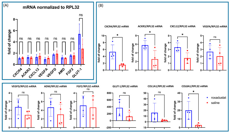
Methods: We evaluated roxadustat's impact on HIF-1 stimulation, cardiac remodeling, and function after MI. Therefore, we analyzed nuclear HIF-1 expression, the mRNA and protein expression of key HIF-1 target genes (RT-PCR, Western blot), inflammatory cell infiltration (immunohistochemistry), and apoptosis (TUNEL staining) 7 days after MI. Additionally, we performed echocardiography in male and female C57BL/6 mice 28 days post-MI.

Results: We found a substantial increase in nuclear HIF-1, associated with an upregulation of HIF-1α target genes like CXCL12/CXCR4/ACKR3 at the mRNA and protein levels. Roxadustat increased the proportion of myocardial reparative M2 CD206+ cells, suggesting beneficial alterations in immune cell migration and a trend towards reduced apoptosis. Echocardiography showed that roxadustat treatment significantly preserved ejection fraction and attenuated subsequent ventricular dilatation, thereby reducing adverse remodeling.

Conclusions: Our findings suggest that roxadustat is a promising clinically approved treatment option to preserve myocardial function by attenuating adverse remodeling.

Keywords: acute coronary syndrome; adverse remodeling; apoptosis; cardiomyopathy; chemokines; fibrosis; hypoxia inducible factor-1; inflammation; prolyl hydroxylase inhibitor.


Conflict of interest statement

Santhosh Kumar Ghadge was involved in research work at the Medical University Innsbruck and is now an employee of Valneva SE. Moritz Messner received funding from the “Tiroler Wissenschaftsförderung” with the grant number “TWF–F.18815”.

Figures

Figure 1
Figure 1
Western blots of the dosage optimization experiment in HUVEC cells. (A) Nuclear fraction of HIF-1α and TBP for loading control. (B) Nuclear HIF-1α normalized to TATA-binding protein (TBP) suggests a dose optimum at 100 µM. (C) Nuclear HIF-1α and TBP for loading control. (D) Nuclear HIF-1α expression shows a steady rise over time and reaches its maximum after 24 h.
Figure 2
Figure 2
(A) Experimental design. (B) In vivo time course of whole heart tissue samples 1–24 h after the application of 50 mg/kg roxadustat. (C) Nuclear HIF-1α expression increases notably after 6 h and further rises until 24 h. Experiments were repeated 3 times.
Figure 3
Figure 3
Quantitative PCR of selected HIF-1 regulated genes seven days after roxadustat treatment (n = 4) compared to placebo controls (n = 4) normalized to RPL32. (A) We did not observe any changes in mRNA expression after seven days of roxadustat treatment in the absence of myocardial infarction. (B) Seven days after MI and roxadustat treatment, we observed a substantial upregulation of CXCL12, CXCR4, ACKR3, and COL8A1 mRNA. All data represent mean ± SD. p-values: ns > 0.05; * ≤ 0.05.
Figure 4
Figure 4
Western blots of whole heart tissue samples 7 days after LAD ligation. (A) Roxadustat-treated (E1–3; n = 3) vs. placebo-treated control (C1–3; n = 3) mice protein expression of ACKR3, CXCR4, CXCL12, and glyceraldehyde-3-phosphate dehydrogenase (GAPDH) as a loading control. (B) ACKR3 and CXCR4 were significantly higher expressed in the roxadustat-treated group, while CXCL12 was only slightly elevated. All data represent mean ± SD. p-values: ns > 0.05; * ≤ 0.05.
Figure 5
Figure 5
(A) M2 macrophage CD 206+ staining of heart tissue from roxadustat-treated and placebo mice, with a heat map indicating expression levels. (B) The accompanying bar graphs compare the quantities of immune cell markers CD45, CD68, and CD206 between the roxadusat-treated and placebo-treated groups, reflecting changes in leukocyte infiltration and macrophage activity post-treatment. All data represent mean ± SD. p-values: ns > 0.05; * ≤ 0.05.
Figure 6
Figure 6
(A) Immunofluorescence images of roxadustat-(top left) and saline-(top right) treated sections were fixed and stained for DAPI (blue) or TUNEL (red). (B) The percentage difference in apoptotic index between the roxadustat and saline groups did not exhibit a significant difference. All data represent mean ± SD. p-values: ns > 0.05.
Figure 7
Figure 7
Echocardiographic measurements. (A) M-Mode of the LV cavity in the parasternal long axis view on a mid-ventricular level (blue = roxadustat-treated mice; red = placebo mice). (B) Echocardiographic measurements comparing roxadustat-treated (n = 10; blue) and saline-treated infarcted mice (n = 10; red). All data represent mean ± SD. p-values: ns > 0.05; * ≤ 0.05 ** ≤ 0.01; *** ≤ 0.001.
Figure 8
Figure 8
(A) Picro-Sirus Red-stained heart sections. (B) Bar graphs representing infarct area and LV wall diameter (n = 10; blue bars) and placebo-treated mice (n = 10; red bars). All data represent mean ± SD; ns not significant.

References

    1. McDonagh T.A., Metra M., Adamo M., Gardner R.S., Baumbach A., Böhm M., Burri H., Butler J., Čelutkienė J., Chioncel O., et al. 2021 ESC Guidelines for the diagnosis and treatment of acute and chronic heart failure. Eur. Heart J. 2021;42:3599–3726. doi: 10.1093/eurheartj/ehab368. - DOI - PubMed
    1. Schofield C.J., Ratcliffe P.J. Oxygen sensing by HIF hydroxylases. Nat. Rev. Mol. Cell Biol. 2004;5:343–354. doi: 10.1038/nrm1366. - DOI - PubMed
    1. Yfantis A., Mylonis I., Chachami G., Nikolaidis M., Amoutzias G.D., Paraskeva E., Simos G. Transcriptional Response to Hypoxia: The Role of HIF-1-Associated Co-Regulators. Cells. 2023;12:798. doi: 10.3390/cells12050798. - DOI - PMC - PubMed
    1. Semenza G.L. Hypoxia-inducible factor 1: Control of oxygen homeostasis in health and disease. Pediatr. Res. 2001;49:614–617. doi: 10.1203/00006450-200105000-00002. - DOI - PubMed
    1. Myllyharju J., Schipani E. Extracellular matrix genes as hypoxia-inducible targets. Cell Tissue Res. 2010;339:19–29. doi: 10.1007/s00441-009-0841-7. - DOI - PMC - PubMed

Publication types

MeSH terms

Grants and funding

LinkOut - more resources