. 2024 Jun 26;13(13):1108.
doi: 10.3390/cells13131108.

Bromodomain Protein Inhibition Protects β-Cells from Cytokine-Induced Death and Dysfunction via Antagonism of NF-κB Pathway

Affiliations

Bromodomain Protein Inhibition Protects β-Cells from Cytokine-Induced Death and Dysfunction via Antagonism of NF-κB Pathway

Vinny Negi et al. Cells. .

Abstract

Cytokine-induced β-cell apoptosis is a major pathogenic mechanism in type 1 diabetes (T1D). Despite significant advances in understanding its underlying mechanisms, few drugs have been translated to protect β-cells in T1D. Epigenetic modulators such as bromodomain-containing BET (bromo- and extra-terminal) proteins are important regulators of immune responses. Pre-clinical studies have demonstrated a protective effect of BET inhibitors in an NOD (non-obese diabetes) mouse model of T1D. However, the effect of BET protein inhibition on β-cell function in response to cytokines is unknown. Here, we demonstrate that I-BET, a BET protein inhibitor, protected β-cells from cytokine-induced dysfunction and death. In vivo administration of I-BET to mice exposed to low-dose STZ (streptozotocin), a model of T1D, significantly reduced β-cell apoptosis, suggesting a cytoprotective function. Mechanistically, I-BET treatment inhibited cytokine-induced NF-kB signaling and enhanced FOXO1-mediated anti-oxidant response in β-cells. RNA-Seq analysis revealed that I-BET treatment also suppressed pathways involved in apoptosis while maintaining the expression of genes critical for β-cell function, such as Pdx1 and Ins1. Taken together, this study demonstrates that I-BET is effective in protecting β-cells from cytokine-induced dysfunction and apoptosis, and targeting BET proteins could have potential therapeutic value in preserving β-cell functional mass in T1D.

Keywords: Brd4; I-BET; NF-kB; STZ; apoptosis; bromodomain; cytokines; diabetes; inflammation; insulin; islet; β-cells.


Conflict of interest statement

There are no conflicts of interest.

Figures

Figure 1
Figure 1
I-BET protects against cytokine-induced β-cell dysfunction. INS-1 cells were pre-treated with I-BET or VC (vehicle control) for 48 h, and then a cytokine cocktail (CC) was added for another 8 h. (AE) Gene expressions by RT-qPCR of (A) Ins1, (B) Ins2, (C) Pdx1, (D) MafA, and (E) Pax6 are shown after normalization to the housekeeping gene as a fold change over VC. (FI) The protein level by Western blotting and the quantification by densitometry of 4 independent experiments are shown for Pdx1 (F,G) and MafA (H,I) and represented as a fold change over VC. α-Tubulin was used as a loading control. (J,K) The secreted insulin from INS-1 cells in basal 2.8 mM glucose (J) and after incubation in indicated glucose concentrations (K) is shown. Insulin secretion is represented as an insulin stimulation index (K) as a fold change over the respective levels from the basal 2.8 mM glucose. (L) Insulin content is measured in INS-1 cell lysates normalized to cellular DNA. The data are represented as mean ± sem (n = 3–7) with at least three independent experiments. Statistical significance was calculated using one-way ANOVA; *** p < 0.001, ** p < 0.01, * p < 0.05.
Figure 2
Figure 2
I-BET protects against cytokine-induced β-cell apoptosis in vitro. (A,B) INS-1 cells pre-treated with I-BET or VC (vehicle control) for 48 h and then exposed to a cytokine cocktail for another 24 h were evaluated by PI and AnnexinV staining by flow cytometry with appropriate controls. The representative dot plot is shown in (A) for the four groups, and the quantitation of early (Annexin+) and late (Annexin+ PI+) apoptotic cells from three independent experiments is shown in (B). (C,D) The protein level of caspase-3 is shown by Western blotting, and the quantification by densitometry of 4 independent experiments is represented as a fold change over VC. α-Tubulin was used as a loading control. The data are described as mean ± sem (n = 3–4) with at least three independent experiments. Statistical significance was calculated using two-way (B) or one-way ANOVA (D); *** p < 0.001, ** p < 0.01, * p < 0.05.
Figure 3
Figure 3
I-BET mediates its effect by antagonizing NF-kB pathway activation. (A,B) The expression of NF-kB target genes—Myc and Xiap—by RT-qPCR shown after normalization to the housekeeping gene as a fold change over VC. (C–I) The phospho-protein and protein level by Western blotting and the quantification by densitometry of 3-4 independent experiments are shown for p65 (C,D), IKKA/B (E,F), and IκBα (GI). α-Tubulin was used as a loading control. Quantitation from densitometry is represented as a fold change over VC. The data are described as mean ± sem (n = 3–4), with at least three independent experiments. Statistical significance was calculated using one-way ANOVA; *** p < 0.001, ** p < 0.01, * p <0.05.
Figure 4
Figure 4
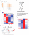
Global transcriptomic changes after I-BET rescues the cytokine-induced changes in INS-1 cells. RNA-Seq analysis from INS-1 cells treated with I-BET or vehicle control (VC) under short (8 h) exposure to the cytokine cocktail (CC). (A) The top 10 enriched pathways of differentially expressed genes (DEGs) between CCIB and CCVC, as determined by IPA. The colors orange and grey depict positive and zero z-scores, respectively. The shades of orange represent stronger correlation with deeper color. (B) The Venn diagram illustrates the genes that were reverted by I-BET in the presence of cytokines. Subsequently, we have the top 5 enriched pathways from those genes as determined by DAVID analysis. (C) A heatmap of pancreatic β-cell function genes that were reverted by I-BET with cytokines. Red and blue indicate up- and down-regulated Z-score-normalized gene expression. (D) The insulin secretion pathway was also among the differentially regulated pathways between CCIB and CCVC. Red and blue represent the up- and down-regulated genes based on their log2 fold change. (E) A heatmap of apoptosis-relevant genes regulated by I-BET, where red and blue indicate up- and down-regulated Z-score-normalized gene expression.
Figure 5
Figure 5
I-BET prevents cytokine-mediated suppression of Foxo1. (A) The expression of Foxo1 mRNA in INS-1 cells was induced by cytokine cocktails (CCs) in 8 h as determined by qRT-PCR and normalized by loading control with respect to VC. (BD) The protein level of phosphorylated Foxo1 and total Foxo1, along with loading control (alpha-tubulin (α-Tub)) is shown in (B). Its respective densitometry is represented in (C,D). (E) The Foxo1 target gene Sod1’s expression was measured in the same condition using qRT-PCR and normalized by loading control and VC. The data are represented as mean ± sem (n = 3–4), with at least three independent experiments. Statistical significance was calculated using one-way ANOVA; *** p < 0.001, ** p < 0.01, * p < 0.05.
Figure 6
Figure 6
I-BET protects against cytokine-induced β-cell apoptosis in vivo. (A) An experimental scheme using the multiple low-dose STZ mouse model was used. (B) Fasting blood glucose level (n = 9 mice/group). (C,D) Representative sections (C) from the pancreas stained for cleaved caspase-3 (green), insulin (red), and DAPI (blue) are shown. The scale bar is 10 µm. The quantitation of cleaved caspase-3 signal from insulin-positive β-cells in each islet is shown (D) with n = 4 mice/group with 2-3 slides for each mouse. The data are represented as mean ± sem. Statistical significance was calculated using two-way ANOVA; *** p < 0.001, ** p < 0.01, * p < 0.05.
Figure 7
Figure 7
I-BET protects against cytokine-induced apoptosis by antagonizing NF-kB signaling. Under inflammation, cytokines bind to their receptors, leading to the phosphorylation of IKKA/B, which in turn leads to the phosphorylation and degradation of IκBα, relieving p65 inhibition. Phospho-p65 then translocates to the nucleus, interacting with BRD proteins and regulating NF-κB target gene transcription, including the repression of β-cell function-related genes such as INS-1, Ins2, and Pdx1, and anti-apoptotic genes (Xiap), and the activation of inflammatory genes such as Myc, leading to reduced insulin secretion, β-cell dysfunction, and ultimately apoptosis. However, when treated with I-BET, the phosphorylation of IKKA/B, IκBα, and p65 is reduced, inhibiting the binding of BRD to p65, indicating an overall decrease in NF-κB pathway activity, reversing the gene expression alterations, improving β-cell function, and reducing apoptosis. Additionally, I-BET-regulated Foxo1 expression and its downstream target Sod1 reduce oxidative stress.

References

    1. Gillespie K.M. Type 1 diabetes: Pathogenesis and prevention. CMAJ. 2006;175:165–170. doi: 10.1503/cmaj.060244. - DOI - PMC - PubMed
    1. Bluestone J.A., Herold K., Eisenbarth G. Genetics, Pathogenesis and clinical interventions in type 1 diabetes. Nature. 2010;464:1293–1300. doi: 10.1038/nature08933. - DOI - PMC - PubMed
    1. Fowler M.J. Microvascular and macrovascular complications of diabetes. Clin. Diabetes. 2008;26:77–82. doi: 10.2337/diaclin.26.2.77. - DOI
    1. Goudy K., Song S., Wasserfall C., Zhang Y.C., Kapturczak M., Muir A., Powers M., Scott-Jorgensen M., Campbell-Thompson M., Crawford J.M., et al. Adeno-associated virus vector-mediated Il-10 gene delivery prevents type 1 diabetes in NOD mice. Proc. Natl. Acad. Sci. USA. 2001;98:13913–13918. doi: 10.1073/pnas.251532298. - DOI - PMC - PubMed
    1. Shomali M. Diabetes treatment in 2025: Can scientific advances keep pace with prevalence? Ther. Adv. Endocrinol. Metab. 2012;3:163–173. doi: 10.1177/2042018812465639. - DOI - PMC - PubMed

Publication types

MeSH terms

LinkOut - more resources