. 2024 Jun 29;13(13):1128.
doi: 10.3390/cells13131128.

Inhibition of Vascular Smooth Muscle Cell Proliferation by ENPP1: The Role of CD73 and the Adenosine Signaling Axis

Affiliations

Inhibition of Vascular Smooth Muscle Cell Proliferation by ENPP1: The Role of CD73 and the Adenosine Signaling Axis

Boris Tchernychev et al. Cells. .

Abstract

The Ectonucleotide Pyrophosphatase/Phosphodiesterase 1 (ENPP1) ectoenzyme regulates vascular intimal proliferation and mineralization of bone and soft tissues. ENPP1 variants cause Generalized Arterial Calcification of Infancy (GACI), a rare genetic disorder characterized by ectopic calcification, intimal proliferation, and stenosis of large- and medium-sized arteries. ENPP1 hydrolyzes extracellular ATP to pyrophosphate (PPi) and AMP. AMP is the precursor of adenosine, which has been implicated in the control of neointimal formation. Herein, we demonstrate that an ENPP1-Fc recombinant therapeutic inhibits proliferation of vascular smooth muscle cells (VSMCs) in vitro and in vivo. Addition of ENPP1 and ATP to cultured VSMCs generated AMP, which was metabolized to adenosine. It also significantly decreased cell proliferation. AMP or adenosine alone inhibited VSMC growth. Inhibition of ecto-5'-nucleotidase CD73 decreased adenosine accumulation and suppressed the anti-proliferative effects of ENPP1/ATP. Addition of AMP increased cAMP synthesis and phosphorylation of VASP at Ser157. This AMP-mediated cAMP increase was abrogated by CD73 inhibitors or by A2aR and A2bR antagonists. Ligation of the carotid artery promoted neointimal hyperplasia in wild-type mice, which was exacerbated in ENPP1-deficient ttw/ttw mice. Prophylactic or therapeutic treatments with ENPP1 significantly reduced intimal hyperplasia not only in ttw/ttw but also in wild-type mice. These findings provide the first insight into the mechanism of the anti-proliferative effect of ENPP1 and broaden its potential therapeutic applications beyond enzyme replacement therapy.

Keywords: neointima; purinergic signaling; vascular smooth muscle cells.


Conflict of interest statement

B.T., D.C., C.S., L.F., K.O., J.H., D.T., D.O. and Y.S. are current employees and stockholders of Inozyme Pharma. Z.C. was an employee of Inozyme Pharma at the time of the study. F.R. is stockholder of Inozyme Pharma and received consulting fees from Inozyme Pharma. The remaining authors declare that they have no conflicts of interest.

Figures
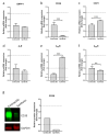
Figure 1
Figure 1
Analysis of ectonucleotidases and adenosine receptors mRNA in contractile and synthetic VSMCs. VSMCs were differentiated towards synthetic or contractile phenotypes. Expression of mRNA encoding ENPP1 (a), ATP/ADP-specific ectonucleotidase CD39 (b), AMP-specific ectonucleotidase CD73 (c), and A1, A2a, A2b adenosine receptors (df) was analyzed using qPCR. Expression of CD39 protein in contractile and synthetic VSMCs (g). Values are presented as the mean ± SD, n = 6, ** p < 0.01, *** p < 0.001 (t-test).
Figure 2
Figure 2
Effect of ENPP1—Fc, AMP, and adenosine (Ado) on VSMCs proliferation. Synthetic VSMCs were starved for 24 h in basal media. Cells were then cultured for 72 h in FBS (5%) containing media in the presence or absence of ENPP1—Fc/ATP (a), 300 μM ATP alone (a), AMP or adenosine (b). Cell proliferation was evaluated by BrdU incorporation. Values are presented as the mean ± SD, n = 8. ** p < 0.01, *** p < 0.001 (one-way ANOVA).
Figure 3
Figure 3
Role of CD73 in antiproliferative effect of ENPP1-Fc/ATP and AMP. Synthetic VSMCs were starved for 24 h in serum-free media. Then cells were cultured in a media supplemented with heat-inactivated FBS (5%) in the presence of AB680 and, either (a) ENPP1-Fc (0.2 μg/mL) and ATP (300 μM) or (b) AMP (300 μM). After 3 days, cell proliferation was evaluated by BrdU incorporation. Values are presented as the mean ± SD, n = 8, ** p < 0.01, *** p < 0.001, (one-way ANOVA).
Figure 4
Figure 4
Metabolism of extracellular AMP by VSMCs. Synthetic VSMCs were incubated in basal media supplemented with 300 μM ATP (a,b) or with ENPP1-Fc (0.2 μg /mL) and ATP (ac) in the presence or absence of CD73 inhibitor AB680 (c). Concentrations of adenosine and inosine in culture media were determined by HPLC. Values are presented as the mean ± SD, n = 6–8.
Figure 5
Figure 5
Effect of extracellular AMP and CD73 inhibition on cAMP levels in VSMCs. Synthetic VSMCs were pretreated for 18 h with vehicle (Veh) or CD73 inhibitor AB680 (1 μM). Cells were washed and stimulated for 4 h with AMP in the presence of vehicle or AB680. Total cAMP was measured using HTRF assay. Values are presented as the mean ± SD, n = 6, *** p < 0.001 (t-test).
Figure 6
Figure 6
Effect of AMP on activation of Gs-coupled adenosine receptors and PKA. Synthetic VSMCs were pretreated for 18 h with either (a) dual A2aR/A2bR antagonist AB928 (0.3 μM) or (b) selective A2aR antagonist SCH-58261 (1 μM) and selective A2bR antagonist PSB-1115 (1 μM). Cells were washed and stimulated for 4 h with AMP in the presence or absence of inhibitors. Total cAMP was measured using HTRF assay. VSMCs were preincubated for 30 min in the basal media supplemented with 0.25% FBS and stimulated for 30 min with AMP in the absence (c) or presence (d) of PKA inhibitor KT5720. Phosphorylation of VASP at Ser157 was analyzed using HTRF assay. Values are presented as the mean ± SD, n = 4–6, ** p < 0.01, *** p < 0.001 (t-test).
Figure 7
Figure 7
Effect of prophylactic treatment with INZ-701 on neointima formation in WT and ttw/ttw mice. Treatment of mice with either vehicle or INZ-701 (10 mg/kg) started 7 days before ligation of left carotid artery. Carotid arteries were collected on day 14th after ligation and fixed on formalin. Morphometric analysis of elastin-stained sections was performed to determine medial (a) and intimal (b) areas, and the intima/media ratio (c). Elastica von Gieson’s staining of carotid arteries cross sections from vehicle or INZ-701-treated WT and ttw/ttw mice (d). Smooth muscle cell proliferation was assessed by immunohistochemical staining of Ki-67 (arrowhead) (e). From outside to inside, external elastic lamina, internal elastic lamina, and luminal boarder (if existing) are circled for better visualization in (d,e). The area between external elastic lamina and internal elastic lamina displays the medial area (#), the area between internal elastic lamina and luminal boarder displays the intimal area (star). Staining of alpha smooth muscle actin (f) Values are presented as the mean ± SD, n = 10–12, * p < 0.05, ** p < 0.01, *** p < 0.001, **** p < 0.0001 (one-way ANOVA).
Figure 8
Figure 8
Effect of therapeutic treatment with INZ-701 on neointima formation in WT and ttw/ttw mice. Treatment of mice with either vehicle or INZ-701 (10 mg/kg) started on day 7 after ligation of left carotid artery. Carotid arteries were collected on day 14th after ligation and fixed in formalin. Morphometric analysis of elastin-stained sections was performed to determine medial (a) and intimal (b) areas, and the intima/media ratio (c). Elastin von Gieson’s staining of carotid arteries cross sections from vehicle- or INZ-701-treated WT and ttw/ttw mice (d). From outside to inside, external elastic lamina, internal elastic lamina, and luminal boarder are circled for better visualization in (d). The area between external elastic lamina and internal elastic lamina displays the medial area (#), the area between internal elastic lamina and luminal boarder displays the intimal area (star). Values are presented as the mean ± SD, n = 8–11, * p < 0.05, ** p < 0.01, *** p < 0.001 (one-way ANOVA).

References

    1. Schermuly R.T., Ghofrani H.A., Wilkins M.R., Grimminger F. Mechanisms of disease: Pulmonary arterial hypertension. Nat. Rev. Cardiol. 2011;8:443–455. doi: 10.1038/nrcardio.2011.87. - DOI - PMC - PubMed
    1. Sakao S., Voelkel N.F., Tatsumi K. The vascular bed in COPD: Pulmonary hypertension and pulmonary vascular alterations. Eur. Respir. Rev. 2014;23:350–355. doi: 10.1183/09059180.00007913. - DOI - PMC - PubMed
    1. Fox B.M., Dorschel K.B., Lawton M.T., Wanebo J.E. Pathophysiology of Vascular Stenosis and Remodeling in Moyamoya Disease. Front. Neurol. 2021;12:661578. doi: 10.3389/fneur.2021.661578. - DOI - PMC - PubMed
    1. Lee T., Roy-Chaudhury P. Advances and new frontiers in the pathophysiology of venous neointimal hyperplasia and dialysis access stenosis. Adv. Chronic Kidney Dis. 2009;16:329–338. doi: 10.1053/j.ackd.2009.06.009. - DOI - PMC - PubMed
    1. Lee T., Chauhan V., Krishnamoorthy M., Wang Y., Arend L., Mistry M.J., El-Khatib M., Banerjee R., Munda R., Roy-Chaudhury P. Severe venous neointimal hyperplasia prior to dialysis access surgery. Nephrol. Dial. Transplant. 2011;26:2264–2270. doi: 10.1093/ndt/gfq733. - DOI - PMC - PubMed

Publication types

MeSH terms

LinkOut - more resources