. 2024 Jul 3;13(13):1141.
doi: 10.3390/cells13131141.

Mild Hyperthermia-Induced Thermogenesis in the Endoplasmic Reticulum Defines Stress Response Mechanisms

Affiliations

Mild Hyperthermia-Induced Thermogenesis in the Endoplasmic Reticulum Defines Stress Response Mechanisms

Barbara Dukic et al. Cells. .

Abstract

Previous studies reported that a mild, non-protein-denaturing, fever-like temperature increase induced the unfolded protein response (UPR) in mammalian cells. Our dSTORM super-resolution microscopy experiments revealed that the master regulator of the UPR, the IRE1 (inositol-requiring enzyme 1) protein, is clustered as a result of UPR activation in a human osteosarcoma cell line (U2OS) upon mild heat stress. Using ER thermo yellow, a temperature-sensitive fluorescent probe targeted to the endoplasmic reticulum (ER), we detected significant intracellular thermogenesis in mouse embryonic fibroblast (MEF) cells. Temperatures reached at least 8 °C higher than the external environment (40 °C), resulting in exceptionally high ER temperatures similar to those previously described for mitochondria. Mild heat-induced thermogenesis in the ER of MEF cells was likely due to the uncoupling of the Ca2+/ATPase (SERCA) pump. The high ER temperatures initiated a pronounced cytosolic heat-shock response in MEF cells, which was significantly lower in U2OS cells in which both the ER thermogenesis and SERCA pump uncoupling were absent. Our results suggest that depending on intrinsic cellular properties, mild hyperthermia-induced intracellular thermogenesis defines the cellular response mechanism and determines the outcome of hyperthermic stress.

Keywords: RT-qPCR; flow cytometry; fluorescence microscopy; heat-shock response; human osteosarcoma cells; mild heat; mouse embryonic fibroblast cells; organelle temperature; spectrofluorometry; super-resolution microscopy; thermogenesis; unfolded protein response.


Conflict of interest statement

The authors declare no conflicts of interest.

Figures
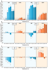
Figure 1
Figure 1
Relative gene expression changes (ddCT) in MEF and U2OS cells in response to heat treatment. The cells were subjected to different temperatures (39 °C, 40 °C, 41 °C, 42 °C, 43 °C, 44 °C) for 1 h or left untreated at 37 °C. The relative expression of different (A) heat-shock protein (HSP) and (B,C) unfolded protein response (UPR) genes was studied using RT-qPCR (n = 3 biological repetitions). Student’s t-test was used for statistical comparisons. Data represent mean ± SD, n = 3, * p < 0.05 compared to 37 °C.
Figure 2
Figure 2
Clustering of the IRE1 transmembrane protein in fixed U2OS IRE1-mEOS cells upon heat treatment (1 h at 40 °C or 42 °C). (A) Size distribution of IRE1 clusters in non-treated (37 °C) and heat-treated (40° or 42 °C) U2OS cells. The Kolmogorov–Smirnov test was performed to analyze the equality of distributions. Samples treated at 40° or 42 °C differed from the control (37 °C) significantly (p < 0.05). Tunicamycin (TM) treatment (5 µg/mL, 1 h) was used as the positive control. (B) Representative 2D image of clusters formed by localizations of IRE1 molecules. Clusters resulting from DBSCAN analysis are marked with different but random colors (see Figure S4).
Figure 3
Figure 3
Flow cytometric determination of the level of XBP1 (mNeonGreen) protein in heat-stressed U2OS cells. Cells were heat-stressed at 40 °C or 42 °C for one hour, followed by six hours of recovery at 37 °C. The Kolmogorov–Smirnov test was performed to analyze the equality of distributions. Samples treated at 40 or 42 °C differed from the control (37 °C) significantly (p < 0.05).
Figure 4
Figure 4
Effect of mild hyperthermia on the endoplasmic reticulum temperature in MEF and U2OS cells. (A) Changes in F/F0 ratio in live, fixed, and 1 µM thapsigargin-pretreated live cells upon 40 °C heat treatment. The average fluorescence intensity values of the heat-treated sample, F, were normalized with the average intensity values of the sample at 37 °C before the heat treatment, F0, by calculating the F/F0 ratio. For F/F0 = 1, there was no change in ER temperature. F/F0 < 1 indicated increased ER temperature, and F/F0 > 1 indicated decreased ER temperature. Studen’s t-test was used for statistical comparisons. Data are mean ± SD, n = 1000, * p < 0.05 compared to fixed cells. (B) Overlay of fluorescence images (blue, Hoechst 33342; yellow, ER thermo yellow) of MEF cells at 37 °C. (C) Calibration curve calculated from the intensity changes in fixed cells (n = 10 cells).
Figure 5
Figure 5
Effect of mild heat on endoplasmic reticulum Ca2+ levels. Flow cytometric analysis of endoplasmic reticulum Ca2+ levels in Mag-fluo-4-labeled, non-treated (37 °C), heat-treated (40 °C), and TG-pretreated (1 µM, 1 h) (A) MEF and (B) U2OS cells. The Kolmogorov–Smirnov test was performed to analyze the equality of distributions. MEF samples treated at 40° or 42 °C differed from the control (37 °C) significantly. TG: thapsigargin.
Figure 6
Figure 6
Cell-specific alternative stress response mechanisms. Under mild hyperthermia, U2OS cells implement both the UPR and HSR to maintain membrane and protein homeostasis, while under more severe heat conditions (above 42 °C), a second wave of UPR is initiated to handle hyperthermic damage. In MEF cells, however, as a result of membrane perturbation-initiated thermogenesis (SERCA pump uncoupling), the higher local ER temperatures launch a more robust HSR with a downregulated UPR. Because of the higher internal temperature, the second UPR wave starts a few degrees lower in MEF cells, but in both cell lines, this second phase of the stress response is accompanied by mitigation of HSR.

Similar articles

Cited by

References

    1. Almanza A., Carlesso A., Chintha C., Creedican S., Doultsinos D., Leuzzi B., Luís A., McCarthy N., Montibeller L., More S., et al. Endoplasmic Reticulum Stress Signalling – from Basic Mechanisms to Clinical Applications. FEBS J. 2019;286:241–278. doi: 10.1111/febs.14608. - DOI - PMC - PubMed
    1. Chen X., Shi C., He M., Xiong S., Xia X. Endoplasmic Reticulum Stress: Molecular Mechanism and Therapeutic Targets - Signal Transduction and Targeted Therapy. Signal Transduct. Target. Ther. 2023;8:352. doi: 10.1038/s41392-023-01570-w. - DOI - PMC - PubMed
    1. Ron D., Walter P. Signal Integration in the Endoplasmic Reticulum Unfolded Protein Response. Nat. Rev. Mol. Cell Biol. 2007;8:519–529. doi: 10.1038/nrm2199. - DOI - PubMed
    1. Peksel B., Gombos I., Péter M., Vigh L., Tiszlavicz Á., Brameshuber M., Balogh G., Schütz G.J., Horváth I., Vigh L., et al. Mild Heat Induces a Distinct “Eustress” Response in Chinese Hamster Ovary Cells but Does Not Induce Heat Shock Protein Synthesis. Sci. Rep. 2017;7:15643. doi: 10.1038/s41598-017-15821-8. - DOI - PMC - PubMed
    1. Tiszlavicz Á., Gombos I., Péter M., Hegedűs Z., Hunya Á., Dukic B., Nagy I., Peksel B., Balogh G., Horváth I., et al. Distinct Cellular Tools of Mild Hyperthermia-Induced Acquired Stress Tolerance in Chinese Hamster Ovary Cells. Biomedicines. 2022;10:1172. doi: 10.3390/biomedicines10051172. - DOI - PMC - PubMed

Publication types

Substances

LinkOut - more resources