. 2024 Jul 8;13(13):1161.
doi: 10.3390/cells13131161.

Tumor Necrosis Factor-Alpha Modulates Expression of Genes Involved in Cytokines and Chemokine Pathways in Proliferative Myoblast Cells

Affiliations

Tumor Necrosis Factor-Alpha Modulates Expression of Genes Involved in Cytokines and Chemokine Pathways in Proliferative Myoblast Cells

Angela María Alvarez et al. Cells. .

Abstract

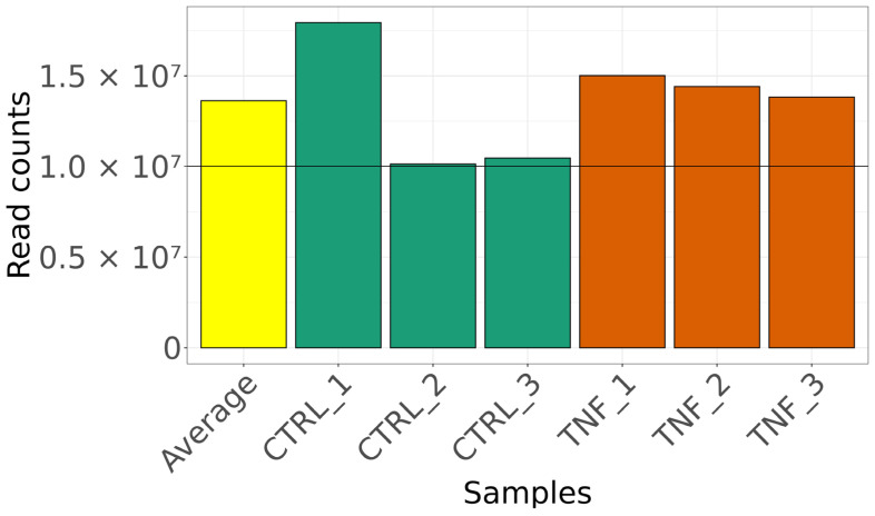
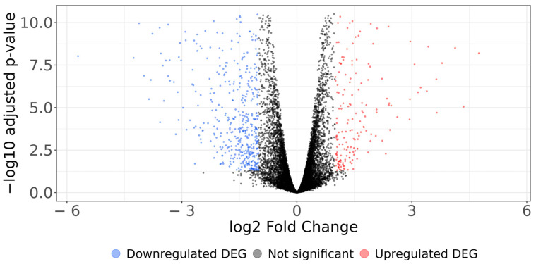
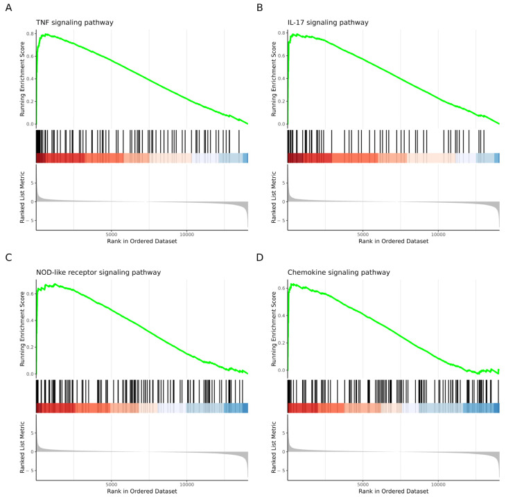
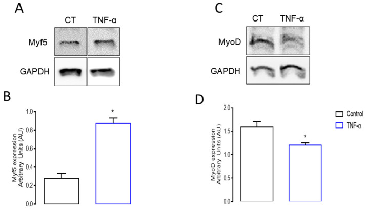
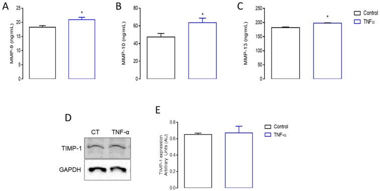
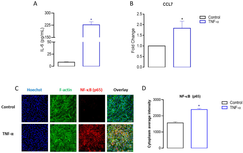
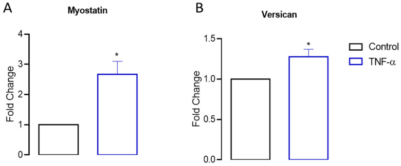
Skeletal muscle regeneration after injury is a complex process involving inflammatory signaling and myoblast activation. Pro-inflammatory cytokines like tumor necrosis factor-alpha (TNF-α) are key mediators, but their effects on gene expression in proliferating myoblasts are unclear. We performed the RNA sequencing of TNF-α treated C2C12 myoblasts to elucidate the signaling pathways and gene networks regulated by TNF-α during myoblast proliferation. The TNF-α (10 ng/mL) treatment of C2C12 cells led to 958 differentially expressed genes compared to the controls. Pathway analysis revealed significant regulation of TNF-α signaling, along with the chemokine and IL-17 pathways. Key upregulated genes included cytokines (e.g., IL-6), chemokines (e.g., CCL7), and matrix metalloproteinases (MMPs). TNF-α increased myogenic factor 5 (Myf5) but decreased MyoD protein levels and stimulated the release of MMP-9, MMP-10, and MMP-13. TNF-α also upregulates versican and myostatin mRNA. Overall, our study demonstrates the TNF-α modulation of distinct gene expression patterns and signaling pathways that likely contribute to enhanced myoblast proliferation while suppressing premature differentiation after muscle injury. Elucidating the mechanisms involved in skeletal muscle regeneration can aid in the development of regeneration-enhancing therapeutics.

Keywords: cytokine; inflammation; muscle regeneration.


Conflict of interest statement

The authors declare no conflicts of interest. The funders had no role in the design of the study; in the collection, analyses, or interpretation of data; in the writing of the manuscript, or in the decision to publish the results.

Figures

Figure 1
Figure 1
Read counts per each library in the RNA-Seq. On the left, colored yellow, is the average of read counts per library. We found 958 differentially expressed genes (DEGs) in samples treated with TNF-α compared to untreated samples, used as control (Supplementary Table S1, Figure 2). Our results agree with other studies reporting that TNF-α modulates the expression of several genes in myoblast cells, using other technologies such as microarray-based gene expression [28,51].
Figure 2
Figure 2
Volcano plot illustrating the differential gene expression between TNF-α treated and untreated samples. The x axis displays the log2 fold change (LFC) or relative abundance, and the y axis presents the adjusted p-value (−log10) obtained through the FDR algorithm. Upregulated genes are red dots on the right side, whereas downregulated genes are green dots on the left, and genes with no significant change are black dots.
Figure 3
Figure 3
GSEA plot showing the most significantly enriched pathways using the KEGG database. The most significantly enriched pathways were (A) the first most significantly enriched pathway was TNF signaling pathway; (B) the second was IL-17 signaling pathway; (C) the third was NOD-like receptor pathway; and (D) the fourth was chemokine signaling pathway.
Figure 4
Figure 4
Protein expression of myogenic regulatory factors Myf5 and MyoD. The expressions of Myf5 and MyoD were assessed by Western blotting in proliferative myoblast cells stimulated with TNF-α for 24 h. Representative blots are presented for both (A) Myf5 and (C) MyoD protein expressions. TNF-α induced an (B) increase in Myf5 expression, while in contrast, (D) MyoD expression decreased after 24 h of TNF-α incubation (* p < 0.05 from unpaired t-test, n = 6).
Figure 5
Figure 5
Matrix metalloproteinases (MMPs) production and tissue inhibitor of metalloproteinase 1 (TIMP-1) expression. MMP-9, 10, and 13 production was measured by ELISA in proliferative myoblast cells stimulated with TNF-α for 24 h. TNF-α treatment resulted in the increased production of (A) MMP-9, (B) MMP-10, and (C) MMP-13. Additionally, the TIMP-1 expression was examined by Western blotting in myoblast cells stimulated with TNF-α for 24 h. Representative blots are shown for TIMP-1 (D), and its densitometric analysis (E) (* p < 0.05 from unpaired t-test, n = 6).
Figure 6
Figure 6
Analysis of the production of IL-6 and the expression of CCL7 and the p65 subunit of NF-KB in C2C12 cells following TNF-α incubation. The levels of IL-6, CCL7, and NF-KB were assessed using ELISA, RT-qPCR, and HCS analysis (20× magnification) in proliferative myoblast cells stimulated with TNF-α for 24 h, as described in the Materials and Methods. TNF-α stimulation led to elevated expression levels of IL-6 (A), CCL7 (B), and the p65 subunit (C,D). Relative changes were compared with the untreated control (* p < 0.05 from unpaired t-test, n = 3 to 4).
Figure 7
Figure 7
Myostatin and versican gene expressions in myoblast C2C12 cells by RT-qPCR assays. Myostatin and versican gene expressions were assessed via RT-qPCR in proliferative myoblast cells stimulated with TNF-α for 24 h, as described in the Materials and Methods. TNF-α stimulation resulted in elevated expression levels of myostatin (A) and versican (B). Relative changes in gene expressions were determined using the (2−ΔΔCt) method and are presented as fold changes compared to the untreated control (* p < 0.05 from unpaired t-test, n = 3).

Similar articles

Cited by

References

    1. Dumont N.A., Bentzinger C.F., Sincennes M.-C., Rudnicki M.A. Satellite Cells and Skeletal Muscle Regeneration. Compr. Physiol. 2015;5:1027–1059. doi: 10.1002/cphy.c140068. - DOI - PubMed
    1. Snijders T., Nederveen J.P., McKay B.R., Joanisse S., Verdijk L.B., van Loon L.J.C., Parise G. Satellite Cells in Human Skeletal Muscle Plasticity. Front. Physiol. 2015;6:283. doi: 10.3389/fphys.2015.00283. - DOI - PMC - PubMed
    1. Kaczmarek A., Kaczmarek M., Ciałowicz M., Clemente F.M., Wolański P., Badicu G., Murawska-Ciałowicz E. The Role of Satellite Cells in Skeletal Muscle Regeneration-The Effect of Exercise and Age. Biology. 2021;10:1056. doi: 10.3390/biology10101056. - DOI - PMC - PubMed
    1. Giuliani G., Rosina M., Reggio A. Signaling Pathways Regulating the Fate of Fibro/Adipogenic Progenitors (FAPs) in Skeletal Muscle Regeneration and Disease. FEBS J. 2022;289:6484–6517. doi: 10.1111/febs.16080. - DOI - PubMed
    1. Hernández-Hernández O., Ávila-Avilés R.D., Hernández-Hernández J.M. Chromatin Landscape During Skeletal Muscle Differentiation. Front. Genet. 2020;11:578712. doi: 10.3389/fgene.2020.578712. - DOI - PMC - PubMed

Publication types

LinkOut - more resources