Review
. 2024 Sep 26;88(3):e0000624.
doi: 10.1128/mmbr.00006-24. Epub 2024 Jul 12.

Evolutionary trajectory for nuclear functions of ciliary transport complex proteins

Affiliations
Review

Evolutionary trajectory for nuclear functions of ciliary transport complex proteins

Alexander Ewerling et al. Microbiol Mol Biol Rev. .

Abstract

SUMMARYCilia and the nucleus were two defining features of the last eukaryotic common ancestor. In early eukaryotic evolution, these structures evolved through the diversification of a common membrane-coating ancestor, the protocoatomer. While in cilia, the descendants of this protein complex evolved into parts of the intraflagellar transport complexes and BBSome, the nucleus gained its selectivity by recruiting protocoatomer-like proteins to the nuclear envelope to form the selective nuclear pore complexes. Recent studies show a growing number of proteins shared between the proteomes of the respective organelles, and it is currently unknown how ciliary transport proteins could acquire nuclear functions and vice versa. The nuclear functions of ciliary proteins are still observable today and remain relevant for the understanding of the disease mechanisms behind ciliopathies. In this work, we review the evolutionary history of cilia and nucleus and their respective defining proteins and integrate current knowledge into theories for early eukaryotic evolution. We postulate a scenario where both compartments co-evolved and that fits current models of eukaryotic evolution, explaining how ciliary proteins and nucleoporins acquired their dual functions.

Keywords: cell biology; cilia; eukaryogenesis; eukaryotes; evolution; intraflagellar transport; last eukaryotic common ancestor; molecular biology; nuclear pore complex; nucleus.


Conflict of interest statement

The authors declare no conflict of interest.

Figures

Fig 1
Fig 1
Overview of a eukaryotic cilium and intraflagellar transport machinery. Cargo is transported from the proximal basal body along the ciliary axoneme to the tip, where cargo is released, the IFT train is re-structured, and sent back toward the basal body.
Fig 2
Fig 2
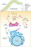
Overview of protocoatomer-derived vesicle coats and their field of action. While parts of the IFT/BBSome are derived from protocoatomer, they are not vesicle coats sensu stricto. They do, however, associate with membranes for cargo delivery.
Fig 3
Fig 3
Structure of a nuclear pore complex. NPCs have eightfold radial symmetry. The NPC is anchored to the NE membrane by transmembrane nucleoporins (membrane ring). Membrane, inner, and outer rings associate with each other to form a barrel-like structure with a central pore. Cargoes enter the nuclear pore at the docking site and are transported by electro- and hydrostatic interactions with phenylalanine-glycine residues. FG residues convey selective transport through the nuclear pore. Cargo complexes leave the pore through the nuclear basket. Transport into the cytoplasm is analogous, with cargoes exiting from the export platforms. Based on reference (49).
Fig 4
Fig 4
(A) Outside-in, (B) inside-out, and (C) endosymbiont-borne models for the generation of endomembrane organelles. (A) Crypts formed by plasma membrane invaginations gradually form a network inside the archaeal cell, increasing the surface and eventually encasing the genetic information. An epibiotic symbiont could be incorporated into the host’s cell body by a larger crypt. Note that numerous membrane fusion events would be needed for membrane scission from the plasma membrane and formation of a separate NE. (B) Protrusions from the archaeal host reach into the extracellular space, making contact with other archaea and possibly symbiotic bacteria [see references (127, 212)]. The tips could then enlarge, engulfing epibiotic bacteria, until contacts with other protrusions would lead to membrane fusion. This would result in lamellar membrane-bounded inclusions in the cytoplasm that would later become the NE and ER. (C) Before the formation of endomembrane organelles, the future endosymbiont is incorporated into the host’s cytoplasm. Constant shedding of outer membrane vesicles (OMVs) from the symbiont would then gradually replace the host’s own plasma membrane. In the cytoplasm, these OMVs could produce GA- and ER-like organelles by fusion. Based on references (12, 209–211).
Fig 5
Fig 5
Sequence of events leading to the ER, GA, cilium, and NE. In the post-FECA cell, the initial protocoatomer likely facilitated directed delivery from the plasma membrane to a proto-ER and vice versa. DNA was freely accessible due to a lack of NE structure. With the enrichment of receptors on a specific patch of the plasma membrane, the cell developed a primitive membrane protrusion, the proto-cilium, that further enhanced signaling capabilities. Pre-existing signaling cascades were inherited, novel pathways established, and regulated via intermediate regulatory levels. With the paralogous expansion of the protocoatomer, the cell was able to differentiate between inward and outward transport and to establish an intermediate compartment for sorting, the proto-GA. The type I coats used for inward transport could also be co-opted by the developing proto-cilium. After further diversification of type I and type II coats, the NE could form with primitive pores, limiting free diffusion of “ciliary” signaling effector proteins into the forming NE. However, some of the ciliary proteins still could enter (either due to the size or presence of advantageous protein structures), while others relied on intermediate messengers that could. At the time the NE with NPCs was fully formed, some signaling pathways must have been lost. Some pathways made use of other proteins that could enter, and others retained their nuclear functions fully.

Similar articles

References

    1. Carvalho-Santos Z, Azimzadeh J, Pereira-Leal JB, Bettencourt-Dias M. 2011. Tracing the origins of centrioles, cilia, and flagella. J Cell Biol 194:165–175. doi: 10.1083/jcb.201011152 - DOI - PMC - PubMed
    1. Lindemann CB. 2022. The flagellar germ-line hypothesis: how flagellate and ciliate gametes significantly shaped the evolution of organismal complexity. Bioessays 44:e2100143. doi: 10.1002/bies.202100143 - DOI - PubMed
    1. Villeneuve AM, Hillers KJ. 2001. Whence meiosis? Cell 106:647–650. doi: 10.1016/s0092-8674(01)00500-1 - DOI - PubMed
    1. Loidl J. 2016. Conservation and variability of meiosis across the eukaryotes. Annu Rev Genet 50:293–316. doi: 10.1146/annurev-genet-120215-035100 - DOI - PubMed
    1. Bremer N, Tria FDK, Skejo J, Garg SG, Martin WF. 2022. Ancestral state reconstructions trace mitochondria but not phagocytosis to the last eukaryotic common ancestor. Genome Biol Evol 14:evac079. doi: 10.1093/gbe/evac079 - DOI - PMC - PubMed

Substances

LinkOut - more resources