. 2024 Jul 12;19(7):e0306611.
doi: 10.1371/journal.pone.0306611. eCollection 2024.

Octopamine is required for successful reproduction in the classical insect model, Rhodnius prolixus

Affiliations

Octopamine is required for successful reproduction in the classical insect model, Rhodnius prolixus

Jimena Leyria et al. PLoS One. .

Abstract

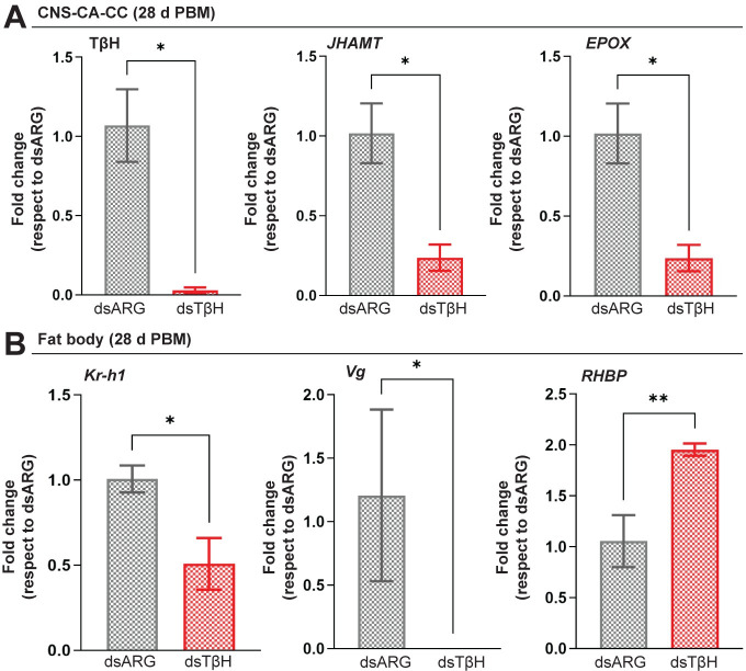
In insects, biogenic amines function as neurotransmitters, neuromodulators, and neurohormones, influencing various behaviors, including those related to reproduction such as response to sex pheromones, oogenesis, oviposition, courtship, and mating. Octopamine (OA), an analog of the vertebrate norepinephrine, is synthesized from the biogenic amine tyramine by the enzyme tyramine β-hydroxylase (TβH). Here, we investigate the mechanisms and target genes underlying the role of OA in successful reproduction in females of Rhodnius prolixus, a vector of Chagas disease, by downregulating TβH mRNA expression (thereby reducing OA content) using RNA interference (RNAi), and in vivo and ex vivo application of OA. Injection of females with dsTβH impairs successful reproduction at least in part, by decreasing the transcript expression of enzymes involved in juvenile hormone biosynthesis, the primary hormone for oogenesis in R. prolixus, thereby interfering with oogenesis, ovulation and oviposition. This study offers valuable insights into the involvement of OA for successful reproduction in R. prolixus females. Understanding the reproductive biology of R. prolixus is crucial in a medical context for controlling the spread of the disease.


Conflict of interest statement

The authors have declared that no competing interests exist.

Figures

Fig 1
Fig 1. Identification of TβH and its transcript expression.
(A) Phylogenetic relationship of TβH in insects. The evolutionary history was inferred by using the Maximum Likelihood method. The tree with the highest log likelihood (-15636.19) is shown. The tree is drawn to scale, with branch lengths measured in the number of substitutions per site. Protein sequences are labelled by species and order name. GenBank accession number: Aedes aegypti, AAEL010856; Aedes albopictus, AALC636_034546; Apis mellifera, NP_001071292.1; Bombyx mori, NP_001243923.1; Cimex lectularius, XP_014250928.1; Culex quinquefasciatus, CQUJHB002793; Drosophila melanogaster, FBgn0010329; Gryllus bimaculatus, BAO52001.1; Musca domestica, MDOA006509; Nilaparvata lugens, XP_039288853.1; Orussus abietinus, XP_012275259.1; Periplaneta americana, AFO63080.1; Schistocerca gregaria, XP_049845783.1; Tribolium castaneum, XP_974169.1; Vespula pensylvanica, XP_043684637.1; Vollenhovia emeryi, XP_011883289.1; Rhodnius tyrosine 3-monooxygenase like (RPRC007034)-. (B) Distribution of TβH transcript in tissues from unfed virgin female R. prolixus. RT-qPCR was used to quantify transcript levels, and data analysis was performed using the 2−ΔCt method. Relative expression values on the y-axes were calculated by geometric averaging of reference genes Rp49 and actin. Data are presented as mean ± SEM (n = 4–5, where each n represents a pool of tissues from 3 insects). **** p < 0.0001 (One-way ANOVA and Tukey’s test as the post hoc test).
Fig 2
Fig 2. Effect of dsRNA treatment at 6 d post blood meal.
(A) Tyrosine decarboxylase (TDC) converts tyrosine into tyramine, which is then converted to octopamine (OA) by tyramine beta-hydroxylase (TβH). TβH transcript downregulation is a mechanism for decreasing OA levels. (B) TβH mRNA expression in the CNS-CC-CA complex of dsTβH-injected mated females at 6 d post blood meal (6 d PBM). Transcript levels were quantified using RT-qPCR and analyzed by the 2-ΔΔCt method. The y axis represents the fold change in expression relative to control (dsARG, value ∼ 1) obtained via geometric averaging using Rp49 and actin as reference genes. The results are shown as the mean ± SEM (n = 7–8, where each n represents an individual tissue from 1 insect). **** p < 0.0001 (Student’s t-test). (C) Upper panel shows representative images of the reproductive system from dsRNA-injected insects 6 d PBM. Note the pink color of oocytes in the controls (dsARG) and white color of oocytes in the dsTβH-injected insects. Lower panels are a higher magnification of representative images which reveal eggs retained in the calyx following the knockdown of TβH. n = 10–15 females.
Fig 3
Fig 3. Egg production in TβH-deficient females.
(A) Average cumulative eggs laid per mated female throughout 18 d post-blood meal (d PBM) after dsARG and dsTβH injections. The results are shown as the mean ± SEM (n = 10 to 15 females). dsARG vs. dsTβH: * p < 0.05; *** p < 0.001 (repeated-measures two-way ANOVA followed by the post-hoc Bonferroni’s test). (B) Representative images showing egg phenotype (left) and egg volume (right) of dsARG- and dsTβH-injected R. prolixus females. The results are shown as the mean ± SEM (n = 15 eggs). **** p < 0.0001 (Student’s t-test). (C) Total cumulative eggs laid per female over 28 d PBM (left) and diagram illustrating the morphology of the ovarioles and representative images showing the female reproductive system morphology (right) of dsRNA-injected females. Note the ovarioles with smaller size of tropharium and terminal oocytes after dsTβH treatment (n = 15–20 females). **** p < 0.0001 (Student’s t-test). (D) Percentage of hatching with respect to total eggs laid after dsRNA treatment (n = 15–20 females). **** p < 0.0001 (Student’s t-test).
Fig 4
Fig 4. Effect of dsRNA treatment on JH biosynthetic enzymes and JH-response genes.
(A) In the CNS-CC-CA, JHAMT and Epox are involved in JHSB3 production, which is released into the circulation to act on the fat body (FB). In the FB, Kr-h1 and Vg are classic JH-response genes. Vg and other yolk protein precursors are then released into the circulation and accumulate in the oocytes by endocytic receptors VgR and LpR. (B) JHAMT and Epox mRNA expression in the CNS-CC-CA complex is decreased in dsTβH-injected mated females at 6 d post blood meal (6 d PBM). (C) Kr-h1, Vg and RHBP mRNA expression in the fat body of dsARG- and dsTβH-injected mated females. Note that Kr-h1 and Vg expression is decreased in dsTβH-injected females. (D) Vg, VgR and LpR mRNA expression in the ovaries of dsARG- and dsTβH-injected mated females. dsTβH injection results in a reduction in Vg expression. Transcript levels were quantified using RT-qPCR and analyzed by the 2-ΔΔCt method. For C and D, the y axes represent the fold change in expression relative to control (dsARG, value ∼ 1) obtained via geometric averaging using Rp49 and actin as reference genes. The results are shown as the mean ± SEM (n = 7–8, where each n represents an individual tissue from 1 insect). * p < 0.05; ** p < 0.01; *** p < 0.001; **** p < 0.0001; ns, not significant (Student’s t-test). Epox, methyl farneseoate epoxidase. JHAMT, juvenile hormone acid O-methyltransferase; Kr-h1, Krüppel homolog 1; LpR, lipophorin receptor; RHBP, Rhodnius heme-binding protein; Vg, vitellogenin; VgR, vitellogenin receptor.
Fig 5
Fig 5. Effect of dsRNA treatment on JH biosynthetic enzymes and JH-response genes 28 days post blood meal (d PBM).
(A) TβH, JHAMT and Epox mRNA expression in the CNS-CC-CA complex of dsARG- and dsTβH-injected females at 28 days post blood meal (28 d PBM). (B) Kr-h1, Vg and RHBP mRNA expression in the fat body of dsARG- and dsTβH-injected insects. RT-qPCR was used to quantify transcript levels, and data analysis was performed using the 2−ΔΔCt method. Relative expression values on the y-axes represent the fold change in expression relative to control (dsARG, value ∼ 1) and were calculated by geometric averaging of reference genes Rp49 and actin. Data are presented as mean ± SEM (n = 7–8, where each n represents an individual tissue from 1 insect). * p < 0.05; ** p < 0.01 (Student’s t-test). CNS-CC-CA, central nervous system-corpora cardiaca-corpora allata; Epox, methyl farneseoate epoxidase; JHAMT, juvenile hormone acid O-methyltransferase; Kr-h1, Krüppel homolog 1; RHBP, Rhodnius heme-binding protein; tyramine β-hydroxylase (TβH); Vg, vitellogenin.
Fig 6
Fig 6. Effect of dsRNA treatment on yolk protein precursors 6 days post blood meal (d PBM).
(A) Total protein content per fat body (mean ± SEM; n = 5–6, where each n represents tissue from 1 insect), SDS-PAGE analysis (10 μg/line, images representative of 3 independent experiments), and western blot (5 μg/line; images representative of 3 independent experiments) after TβH knockdown. (B) Total protein per μL in the hemolymph (mean ± SEM; n = 7–8, where each n represents hemolymph from 1 insect), SDS-PAGE analysis (5 μg/line, images representative of 3 independent experiments), western blot (1 μg/line; images representative of 3 independent experiments), and color of the hemolymph after knockdown of TβH (images representative of n = 5). (C) Total protein content per ovary (mean ± SEM; n = 5–6, where each n represents tissue from 1 insect), SDS-PAGE analysis (10 μg/line, images representative of 3 independent experiments) and western blot (5 μg/line; images representative of 3 independent experiments) after knockdown of TβH. ** p < 0.01; ns, not significant (Student’s t-test). In the SDS-PAGE analyses the molecular weights of the main yolk protein precursor, Vg (or Vt in the ovaries) and RHBP are indicated. RHBP, Rhodnius heme-binding protein; Vg, vitellogenin; Vt, vitellin.
Fig 7
Fig 7. Effect of exogenous octopamine (OA) treatment.
(A) In vivo assays: OA (5 μL of saline containing 10−4 M OA) was injected into unfed virgin females 3 days post ecdysis (d PE), and transcript levels for JHAMT and Epox in the CNS-CC-CA and Kr-h1, Vg and RHBP in the fat body were measured 12 h later. All transcripts were up-regulated by OA except for RHBP. The results are shown as the mean ± SEM (n = 5–6, where each n represents an individual tissue from 1 insect). Transcript expression was quantified using RT-qPCR and analyzed the 2−ΔΔCt method. (B) Ex vivo assays: OA was added to the incubation medium (final concentration: 10−5 M) containing a pool of 3 tissues (CNS-CC-CA from virgin females 3 d PE). After 6 h, JHAMT and Epox mRNA expression was evaluated. The results are shown as the mean ± SEM (n = 4–5, where each n represents a pool of 3 tissues). For A and B, the y axes represent fold change in expression relative to control (saline, value ∼1) obtained via geometric averaging using Rp49 and actin as reference genes. ns, not significant; * p < 0.05; ** p < 0.01; *** p < 0.001 (Student’s t-test). (C) OA treatment on egg laying. OA (5 μL of saline containing 10−4 M of OA) was injected into fed mated females 6 days post blood meal (d PBM), and egg laying pattern was recorded throughout 14 d PBM. The results are shown as the mean ± SEM (n = 10–15 females). ** p < 0.01; *** p < 0.001; **** p < 0.0001 (repeated-measures two-way ANOVA followed by the post-hoc Bonferroni’s test). CNS-CC-CA, central nervous system-corpora cardiaca-corpora allata; Epox, methyl farneseoate epoxidase; JHAMT, juvenile hormone acid O-methyltransferase; Kr-h1, Krüppel homolog 1; OA, octopamine; RHBP, Rhodnius heme-binding protein; Vg, vitellogenin.

Similar articles

References

    1. Orchard I. Octopamine in insects: neurotransmitter, neurohormone, and neuromodulator. Can J Zool. 1992;60: 659–669.
    1. Schoofs L, De Loof A, Van Hiel MB. Neuropeptides as regulators of behavior in insects. Annu Rev Entomol. 2017;62: 35–52. doi: 10.1146/annurev-ento-031616-035500 - DOI - PubMed
    1. Lange AB, Orchard I. Biogenic monoamines in the control of triatomine physiology with emphasis on Rhodnius prolixus. In: Guarneri A, Lorenzo M, editors. Triatominae—The biology of Chagas disease vectors. 1st ed. Cham: Springer Nature Switzerland AG; 2021. p. 145–66.
    1. Barbero F, Mannino G, Casacci LP. The Role of Biogenic Amines in Social Insects: With a Special Focus on Ants. Insects. 2023;14: 386. doi: 10.3390/insects14040386 - DOI - PMC - PubMed
    1. Roeder T. Biochemistry and molecular biology of receptors for biogenic amines in locusts. Microsc Res Tech. 2002;56: 237–247. doi: 10.1002/jemt.10027 - DOI - PubMed

LinkOut - more resources