. 2024 Jul 12;10(28):eadn2091.
doi: 10.1126/sciadv.adn2091. Epub 2024 Jul 12.

Robust reprogramming of glia into neurons by inhibition of Notch signaling and nuclear factor I (NFI) factors in adult mammalian retina

Affiliations

Robust reprogramming of glia into neurons by inhibition of Notch signaling and nuclear factor I (NFI) factors in adult mammalian retina

Nguyet Le et al. Sci Adv. .

Abstract

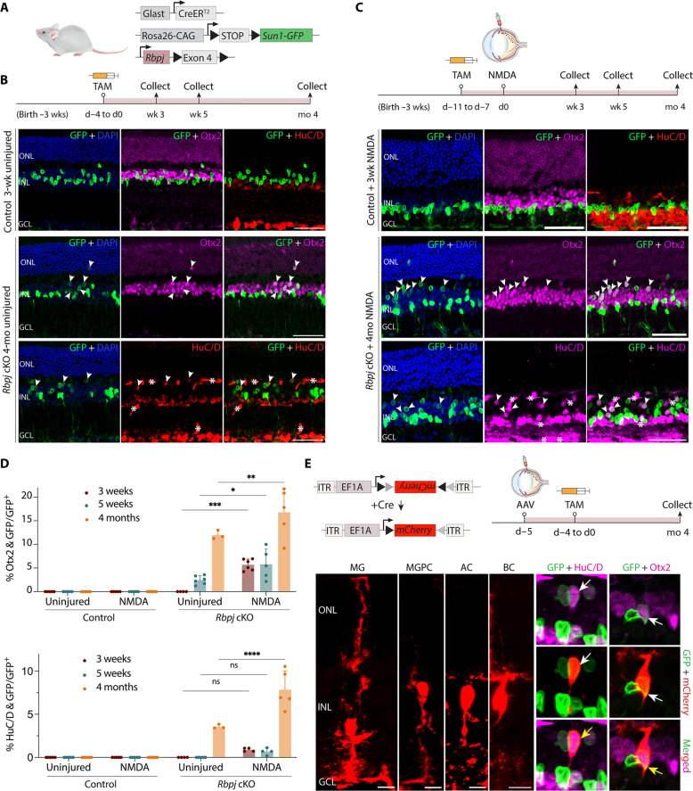
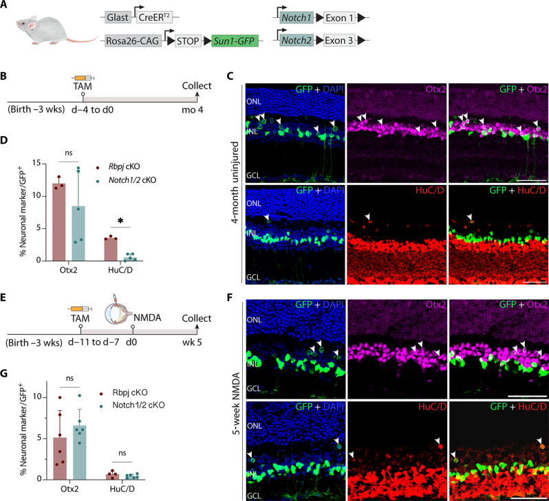
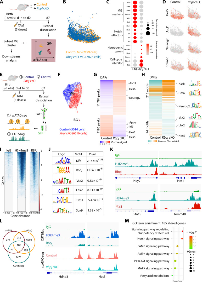
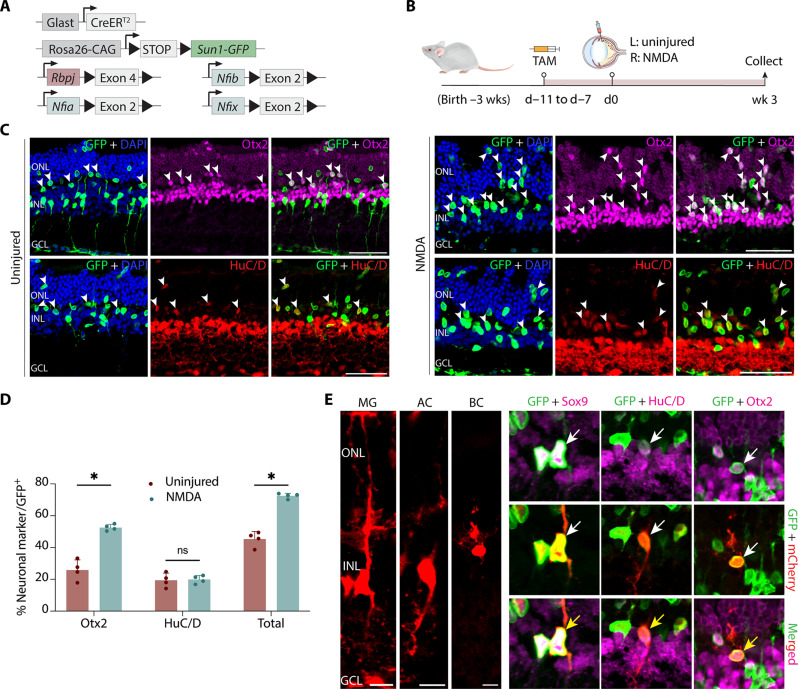
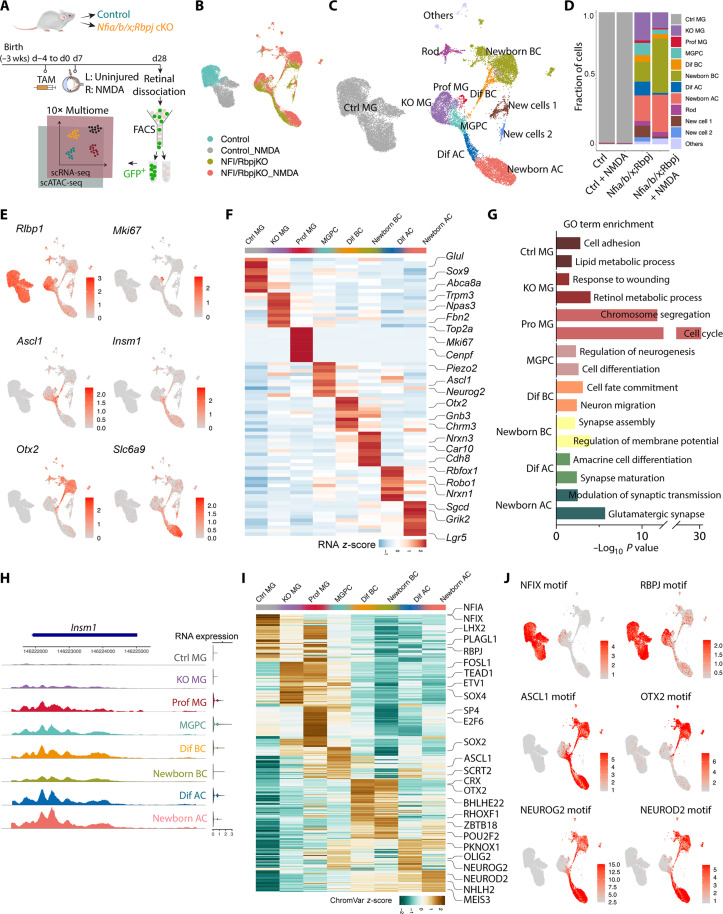
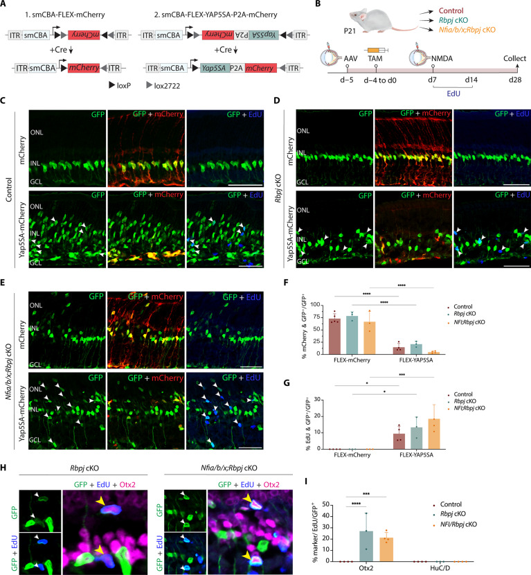
Generation of neurons through direct reprogramming has emerged as a promising therapeutic approach for treating neurodegenerative diseases. In this study, we present an efficient method for reprogramming retinal glial cells into neurons. By suppressing Notch signaling by disrupting either Rbpj or Notch1/2, we induced mature Müller glial cells to reprogram into bipolar- and amacrine-like neurons. We demonstrate that Rbpj directly activates both Notch effector genes and genes specific to mature Müller glia while indirectly repressing expression of neurogenic basic helix-loop-helix (bHLH) factors. Combined loss of function of Rbpj and Nfia/b/x resulted in conversion of nearly all Müller glia to neurons. Last, inducing Müller glial proliferation by overexpression of dominant-active Yap promotes neurogenesis in both Rbpj- and Nfia/b/x/Rbpj-deficient Müller glia. These findings demonstrate that Notch signaling and NFI factors act in parallel to inhibit neurogenic competence in mammalian Müller glia and help clarify potential strategies for regenerative therapies aimed at treating retinal dystrophies.


Figures

Fig. 1.
Fig. 1.. Selective loss of function of Rbpj induces neurogenesis in adult MG with and without retinal injury.
(A) Schematic of the transgenic constructs used to induce deletion of Rbpj specifically in MG. Cre-mediated removal of Exon 4 of Rbpj leads to premature stop codon formation and disrupts the expression of the RBPJ protein. (B and C) Schematic of the experimental workflow and representative immunostaining for green fluorescent protein (GFP), Otx2, and HuC/D in control and Rbpj-deleted retinas without (B) and with (C) NMDA-induced injury. White arrowheads indicate co-labeled GFP-positive and marker-positive cells. GFP-positive MG-derived neuron-like cells often show relatively lower cellular levels of GFP expression than do MG. Asterisks (*) indicate mouse-on-mouse vascular staining. Scale bars, 50 μm. DAPI, 4′,6-diamidino-2-phenylindole. (D) Quantification of mean percentage ± SD of GFP-positive MG-derived neurons expressing either OTX2 or HuC/D. (E) Morphological characterization of MG-derived neurons in Rbpj-deficient retinas using AAV2.7 m8-Ef1a-Flex-mCherry 4 months after TAM treatment. Yellow arrows indicate co-labeled GFP/mCherry/marker-positive cells. Scale bars, 10 μm. Significance was determined via two-way analysis of variance (ANOVA) with Tukey’s multiple comparisons test: *P < 0.05, **P < 0.01, ***P < 0.001, ****P < 0.0001. Each data point was calculated from an individual retina. TAM, tamoxifen; ONL, outer nuclear layer; INL, inner nuclear layer; GCL, ganglion cell layer; MG, Müller glia; MGPC, MG-derived progenitor cell; AC, amacrine cell; BC, bipolar cell; wk/wks, week/weeks; d, day; mo, month.
Fig. 2.
Fig. 2.. Notch1/2 deletion phenocopies effects of Rbpj deletion on MG-derived neurogenesis.
(A) Schematic of the transgenic constructs used to induce deletion of Notch1 and Notch2 specifically in MG. Cre-mediated removal of Exon 1 of Notch1 and Exon 3 of Notch2 leads to premature stop codon formation and disrupts the expression of the NOTCH1/2 proteins. (B) Schematic of the experimental workflow for uninjured retinas. (C and D) Representative images immunolabeled for GFP, OTX2, and HuC/D of Notch1/Notch2-deleted retinas after 4 months after TAM injection, and quantification of mean percentage ± SD of GFP-positive MG-derived neurons expressing either Otx2 or HuC/D. White arrowheads indicate co-labeled GFP-positive and marker-positive cells. (E to G) Schematic of the workflow, representative immunostaining, and quantification for NMDA-injured retinas. Significance was determined via Mann-Whitney U test: *P < 0.05. Each data point was calculated from an individual retina. Scale bars, 50 μm. ns, not signifcant.
Fig. 3.
Fig. 3.. Rbpj deletion in MG down-regulates Notch target genes and activates expression of neurogenic genes.
(A) Schematic of the experimental workflow used to generate scRNA-Seq data from whole retinas of control and Rbpj cKO mice. (B) Uniform manifold approximation and projection (UMAP) of scRNA-seq data showing the clustering of control and Rbpj-deficient MG subsetted from whole retina cell populations. (C) Dot plot showing down-regulation of genes specific to resting MG and Notch-regulated genes, and up-regulation of neurogenic bHLH factors as well as cell cycle inhibitors in Rbpj-deficient MG relative to controls. (D) Feature plots highlighting differential expression of Hes5, Ascl1, Neurog2, and Cdkn1c in control and Rbpj-deficient MG. (E) Schematic of the experimental workflow used to generate scATAC-seq data from control and Rbpj cKO MG and CUT&Tag data from control MG. (F) UMAP plot showing global differences in chromatin accessibility observed by scATAC-seq in control and Rbpj-deficient MG. (G) Differentially accessible chromatin regions (DARs) and nearby genes between Rbpj-deficient and control MG. (H) Differential TF motif enrichments (DMEs) in the DARs between Rbpj-deficient and control MG. (I) CUT&Tag analysis of IgG, H3K4me3, and RBPJ from fluorescence-activated cell sorting (FACS)–isolated GFP-positive MG from GlastCreER;Sun1GFP retinas. (J) TF motifs enriched in RBPJ CUT&Tag samples relative to control. (K) Representative genome tracks for CUT&Tag analysis. (L) Integrative analysis of scRNA-seq, scATAC-seq, and CUT&Tag to identify genes regulated by RBPJ. (M) Gene Ontology (GO) enrichment analysis for genes shared among the three datasets. AMPK, AMP-activated protein kinase; cAMP, cyclic adenosine 3′,5′-monophosphate; MAPK, mitogen-activated protein kinase; PI3K, phosphatidylinositol 3-kinase.
Fig. 4.
Fig. 4.. Simultaneous disruption of Nfia/b/x and Rbpj convert nearly all adult MG into retinal neurons.
(A) Schematic of the transgenic constructs used to induce loss of function of Rbpj and Nfi/a/b/x specifically in MG. (B and C) Schematic of experimental pipeline and representative images of uninjured and NMDA-damaged retinas immunolabeled for GFP, Otx2, and HuC/D. White arrowheads indicate GFP-positive MG-derived neurons expressing neuronal markers Otx2 or HuC/D. (D) Quantification of mean percentage ± SD of GFP-positive MG-derived neurons expressing either Otx2 or HuC/D in uninjured and NMDA-treated retina. (E) Morphological characterization of MG-derived neurons in Nfia/b/x;Rbpj-deficient retinas using AAV2.7 m8-Ef1a-Flex-mCherry 4 weeks after TAM treatment. Yellow arrows indicate co-labeled GFP/mCherry/marker-positive cells. Scale bars, 10 μm. Significance was determined via unpaired t test: *P < 0.05. Scale bars, 50 μm.
Fig. 5.
Fig. 5.. Integrated snRNA/scATAC-seq analysis of control and Nfia/b/x;Rbpj-deficient adult MG and their progeny.
(A) Schematic of the multiomic snRNA/ATAC-seq experimental pipeline. (B) UMAP plot of Multiome (scRNA/ATAC-seq) datasets showing the clustering of control and Nfia/b/x;Rbpj-deficient MG from uninjured and NMDA-treated retinas. (C) UMAP plot showing the identity of cell clusters determined by marker gene expression. (D) Stacked barplots represent the proportion of cells in each cluster across different sample groups. (E) Feature plots highlighting the cluster of MG (Rlbp1), proliferating MG (Mki67), neurogenic MGPC (Ascl1 and Insm1), BCs (Otx2), and ACs (Slc6a9). (F) Heatmap showing the expression of top 10 differentially expressed genes for each cell cluster. (G) GO enrichment for each cell cluster. The x axis indicates the −log10(P value) of the GO term. (H) Representative ATAC peaks and RNA expression level of Insm1 across different cell clusters. (I) Heatmap showing activity of top 50 differential motifs across different cell clusters. (J) Feature plots showing activity of indicated motifs. Prof MG, proliferating MG; Dif BC, differentiating BCs; Dif AC, differentiating ACs.
Fig. 6.
Fig. 6.. Overexpression of dominant active YapS5A stimulates proliferation and enhanced neurogenesis in both Rbpj- and Nfia/b/x/Rbpj-deficient adult MG.
(A) A schematic of Cre-inducible control and Yap5SA FLEX constructs used in the study. (B) Schematic of experimental pipeline. (C to E) Representative immunostaining of GFP, mCherry, and EdU on retinal sections from control, Rbpj cKO, and Nfia/b/x;Rbpj cKO with mCherry control and Yap5SA AAVs. White arrowheads indicate co-labeled GFP-positive and EdU-positive cells. (F) Quantification of AAV transduction efficiency in MG. (G) Quantification of MG co-labeled with EdU. (H) Representative immunostaining of EdU-positive MG-derived neurons expressing BC marker Otx2. Yellow arrowheads indicate EdU/GFP/Otx2 triple-positive cells. (I) Quantification of mean percentage ± SD of EdU-positive MG-derived neurons expressing neuronal markers: Otx2 and HuC/D. Significance was determined via two-way ANOVA with Tukey’s test: *P < 0.05, ***P < 0.001, ****P < 0.0001. Each data point was calculated from an individual retina. Scale bars, 50 μm.

Update of

References

    1. Wan J., Goldman D., Retina regeneration in zebrafish. Curr. Opin. Genet. Dev. 40, 41–47 (2016). - PMC - PubMed
    1. Lahne M., Nagashima M., Hyde D. R., Hitchcock P. F., Reprogramming Müller glia to regenerate retinal neurons. Annu. Rev. Vis. Sci. 6, 171–193 (2020). - PMC - PubMed
    1. Gorsuch R. A., Lahne M., Yarka C. E., Petravick M. E., Li J., Hyde D. R., Sox2 regulates Müller glia reprogramming and proliferation in the regenerating zebrafish retina via Lin28 and Ascl1a. Exp. Eye Res. 161, 174–192 (2017). - PMC - PubMed
    1. Fausett B. V., Gumerson J. D., Goldman D., The proneural basic helix-loop-helix gene ascl1a is required for retina regeneration. J. Neurosci. 28, 1109–1117 (2008). - PMC - PubMed
    1. Jorstad N. L., Wilken M. S., Grimes W. N., Wohl S. G., VandenBosch L. S., Yoshimatsu T., Wong R. O., Rieke F., Reh T. A., Stimulation of functional neuronal regeneration from Müller glia in adult mice. Nature 548, 103–107 (2017). - PMC - PubMed

MeSH terms

Substances