. 2024 Jul 12;10(28):eadn0881.
doi: 10.1126/sciadv.adn0881. Epub 2024 Jul 12.

CAR memory-like NK cells targeting the membrane proximal domain of mesothelin demonstrate promising activity in ovarian cancer

Affiliations

CAR memory-like NK cells targeting the membrane proximal domain of mesothelin demonstrate promising activity in ovarian cancer

Mubin Tarannum et al. Sci Adv. .

Abstract

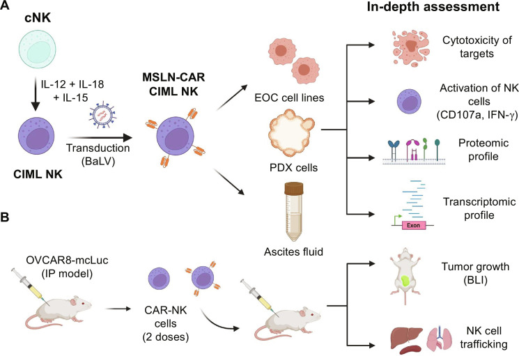
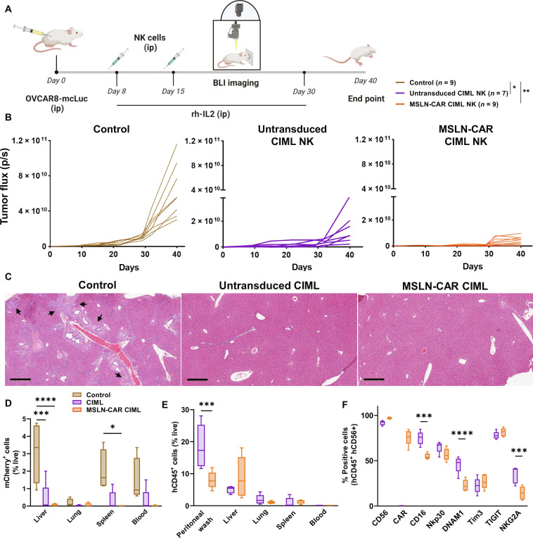
Epithelial ovarian cancer (EOC) remains one of the most lethal gynecological cancers. Cytokine-induced memory-like (CIML) natural killer (NK) cells have shown promising results in preclinical and early-phase clinical trials. In the current study, CIML NK cells demonstrated superior antitumor responses against a panel of EOC cell lines, increased expression of activation receptors, and up-regulation of genes involved in cell cycle/proliferation and down-regulation of inhibitory/suppressive genes. CIML NK cells transduced with a chimeric antigen receptor (CAR) targeting the membrane-proximal domain of mesothelin (MSLN) further improved the antitumor responses against MSLN-expressing EOC cells and patient-derived xenograft tumor cells. CAR arming of the CIML NK cells subtanstially reduced their dysfunction in patient-derived ascites fluid with transcriptomic changes related to altered metabolism and tonic signaling as potential mechanisms. Lastly, the adoptive transfer of MSLN-CAR CIML NK cells demonstrated remarkable inhibition of tumor growth and prevented metastatic spread in xenograft mice, supporting their potential as an effective therapeutic strategy in EOC.


Figures

Fig. 1.
Fig. 1.. Overview of the design, development, and in vitro and in vivo evaluation of MSLN-CAR CIML NK cells.
(A) CIML NK cells were differentiated from cNK by brief (12 to 18 hours) preactivation with IL-12, IL-18, and IL-15 cytokine combination, further armed with MSLN-CAR using Baboon pseudotyped lentivirus (BaLV)–mediated transduction. The activity of MSLN-CAR CIML NK cells was then evaluated against EOC cell lines, PDX tumor cells, and after exposure to patient-derived ascites fluid. After exposure, MSLN-CAR CIML NK cells were evaluated for their antitumor cytotoxicity, activation, proteomic, and transcriptomic changes. (B) In vivo therapeutic efficacy assessment of MSLN-CAR CIML NK cells in an intraperitoneal (IP) model using OVCAR8 cells in nonobese diabetic scid gamma mice (NSG) mice. The graphics in this figure were created with BioRender.com.
Fig. 2.
Fig. 2.. CIML NK cells exhibit superior activity against a panel of EOC cell lines.
(A) Schematic diagram showing the generation of CIML NK cells from cNK cells by 12- to 18-hour activation with the specific cytokine cocktail, 4 to 5 days’ rest and their assessment for cytotoxicity (annexin V/7-AAD), degranulation (CD107a), and IFN-γ production upon 6-hour coculture with EOC cell lines (OVCAR8, SKOV3, OVCAR3, CaOV3, and CaOV4). The graphics were created with BioRender.com. (B) Cytotoxicity of CIML NK cells (purple) versus cNK cells (green) upon coculture with various EOC cell lines at various E:T ratios. (C) Summary of the relative cytotoxicity of CIML NK versus cNK cells at 2:1 E:T ratio. (D) Degranulation (CD107a) and (E) IFN-γ production of CIML NK cells (purple) versus cNK cells (green) upon coculture with various EOC cell lines at 2:1 E:T ratio. Data are represented as means ± SD. Data in (B), (D), and (E) are from NK cells from three independent healthy donors in two technical replicates. Two-way analysis of variance (ANOVA) with donor-matched Sidak’s (B) or Tukey’s [(D) and (E)] multiple comparisons was performed between different groups to determine the statistical difference; ***P ≤ 0.0001, ***P ≤ 0.001, **P ≤ 0.01, and *P ≤ 0.05.
Fig. 3.
Fig. 3.. Proteomic and gene expression differences in the cNK versus CIML NK cells at baseline and gene expression changes in the EOC cells upon coculture with CIML NK cells.
(A) Heatmap showing the expression of key receptors in cNK versus CIML NK cells (three donors) as assessed by an extended flow cytometry NK cell panel. (B) Heatmap (B1) and STRING analysis (B2) of differentially expressed genes in cNK and CIML NK cells (two donors) evaluated by bulk RNA sequencing. (C to E) Volcano plots depicting the differentially expressed genes in EOC cell lines; OVCAR8 (C1), SKOV3 (D1), and OVCAR3 (E1) flow-sorted after 24-hour coculture with CIML NK cells (two donors) at 1:4 E:T ratio. Heatmaps (C2, D2, and E2) and STRING analysis (C3, D3, and E3) of selected genes in EOC cells after coculture with CIML NK cells. Bulk RNA sequencing was performed on flow-sorted EOC cell lines, and data are represented with FDR setup (0.05) and fold change (2).
Fig. 4.
Fig. 4.. Generation and in vitro assessment of MSLN-CAR CIML NK cells.
(A) Schematic representation of the CAR construct design and use of BaLV to transduce CIML NK cells. The graphics were created with BioRender.com. (B) Transduced CIML NK cells stably expressed the CAR construct as assessed by GFP expression on days 5 and 10 after transduction (six donors). (C) Experimental design assessing in vitro antitumor activity of MSLN-CAR CIML NK cells against EOC cell lines (OVCAR8, SKOV3, and OVCAR3). (D) Degranulation (CD107a) and IFN-γ production in untransduced (purple) and MSLN-CAR (orange) CIML NK cells after 6-hour coculture with the EOC cell lines at a 2:1 E:T ratio. (E) Cytotoxicity following long-term (48 hours) coculture of untransduced (purple) versus MSLN-CAR (orange) CIML NK cells with EOC cell lines at various E:T ratios. Data are represented as means ± SD. Data from (D) and (E) show results using NK cells from three independent donors. Two-way ANOVA with donor-matched Tukey’s multiple comparison was performed between different groups to determine the statistical difference; **P ≤ 0.01, and *P ≤ 0.05.
Fig. 5.
Fig. 5.. Assessment of MSLN-CAR CIML NK cell activity against PDX tumor cells.
(A) Schematic representation of PDX cytotoxicity upon coculture with untransduced versus MSLN-CAR CIML NK cells. The graphics were created with BioRender.com. (B) MSLN expression (pink) in the PDX cells from the three models in comparison with isotype (gray). (C) Representative flow plots showing the EpCAM+ apoptotic PDX cells upon coculture with CIML or MSLN-CAR CIML NK cells. (D) Cytotoxicity of untransduced (purple) versus MSLN-CAR CIML NK cells (orange) against the PDX cells after 48-hour coculture at E:T ratios of 5:1 and 10:1. Data are represented as means ± SD. Data in (B) are from five independent healthy NK cell donors. Two-way ANOVA with donor-matched Sidak’s multiple comparison was performed between different groups to determine the statistical difference; ****P ≤ 0.0001.
Fig. 6.
Fig. 6.. Phenotypic, gene expression, and IFN-γ production changes in MSLN-CAR CIML versus untransduced CIML NK cells after exposure to immune-suppressive patient-derived ascites fluid.
(A) Schematic diagram of the study design for the functional assessment of NK cells including IFN-γ secretion, in-depth phenotype, and transcriptomic profile after exposure to EOC patient–derived ascites fluid exposure. The graphics were created with BioRender.com. (B) Cytokine/chemokine analysis of ascites fluid from six different patients with EOC using a 48-marker multiplex panel. (C) IFN-γ production by untransduced (purple) or MSLN-CAR (orange) CIML NK cells after exposure to ascites fluid for 48 hours (checkered pattern) upon coculture with OVCAR8 and SKOV3 target cells for 24 hours at 2:1 E:T ratio and assessed by enzyme-linked immunosorbent assay. Data expressed as fold change of IFN-γ produced from NK cells exposed to ascites versus cells not exposed to ascites. (D) Fold changes in the expression of key activating and inhibitory receptors in untransduced (purple) and MSLN-CAR (orange) CIML NK cells after exposure to ascitic fluid. Data expressed as fold change with the marker expression without ascites fluid normalized to 1.0 (dotted line). (E) Heatmap and STRING analysis of top 20 differentially expressed genes in untransduced and MSLN-CAR CIML NK cells with and without ascitic fluid exposure. Data are represented as means ± SD. Data from (C) and (D) show results using NK cells from three to four independent donors paired with the ascites fluid from three to four patients with EOC, and data from (E) show results from two independent donors paired with ascites fluid from two patients with EOC. Two-way ANOVA with donor-matched Tukey’s multiple comparisons was performed between different groups to determine the statistical difference; ****P ≤ 0.0001, ***P ≤ 0.001, and **P ≤ 0.01.
Fig. 7.
Fig. 7.. MSLN-CAR CIML NK cells exhibit tumor inhibition and prevent tumor cell dissemination in a preclinical xenograft mouse model.
(A) Schematic representation of the therapeutic regimen; NSG mice received OVCAR8-mcLuc (1× 10⁶, ip) followed by PBS (control), untransduced CIML versus MSLN-CAR CIML NK cells (5 × 10⁶, ip) on days 8 and 15 after OVCAR8-mcLuc implantation. Mice were given 75,000 U of recombinant human IL-2 intraperitoneally every other day (to support adoptively transferred NK cells), and tumor burden was assessed by BLI. The graphics were created with BioRender.com. (B) Comparison of tumor burden in mice receiving PBS control (brown), untransduced CIML NK cells (purple), and MSLN-CAR CIML NK cells (orange) and statistical significance shown in (A). (C) Hematoxylin and eosin staining of the liver sections showing the tumor micro-metastasis (shown by arrows) in PBS control versus the treatment groups (scale bar, 200 μm). (D) Comparison of tumor cell spread assessed by flow cytometry of mCherry+ tumor cells in peripheral blood and key organs (lungs, liver, and spleen). (E) Trafficking of the adoptively transferred untransduced CIML and MSLN-CAR CIML NK cells into key organs as assessed by flow cytometry of hCD45+ cells 1 week after adoptive transfer (5 × 10⁶ cells, ip). (F) Expression of key markers in adoptively transferred untransduced or MSLN-CAR CIML NK cells in the peritoneal fluid from the mice in (E). Data are represented as means ± SD. Data in (C) show results compiled from two independent experiments with NK cells from two different donors. Data in (D) are from n = 4 to 5 mice per group. Data in (E) and (F) are from an independent experiment with n = 5 mice per group. Two-way ANOVA Tukey’s multiple comparison was performed between different groups to determine the statistical difference; ****P ≤ 0.0001, ***P ≤ 0.001, **P ≤ 0.01, *P ≤ 0.05.

References

    1. Siegel R. L., Miller K. D., Wagle N. S., Jemal A., Cancer statistics, 2023. CA Cancer J. Clin. 73, 17–48 (2023). - PubMed
    1. Porter R., Matulonis U. A., Immunotherapy for ovarian cancer. Clin. Adv. Hematol. Oncol. 20, 240–253 (2022). - PubMed
    1. Akter S., Rahman M. A., Hasan M. N., Akhter H., Noor P., Islam R., Shin Y., Rahman M. D. H., Gazi M. S., Huda M. N., Nam N. M., Chung J., Han S., Kim B., Kang I., Ha J., Choe W., Choi T. G., Kim S. S., Recent advances in ovarian cancer: Therapeutic strategies, potential biomarkers, and technological improvements. Cells 11, 650 (2022). - PMC - PubMed
    1. Robert C., A decade of immune-checkpoint inhibitors in cancer therapy. Nat. Commun. 11, 3801 (2020). - PMC - PubMed
    1. Shiravand Y., Khodadadi F., Kashani S. M. A., Hosseini-Fard S. R., Hosseini S., Sadeghirad H., Ladwa R., O’Byrne K., Kulasinghe A., Immune checkpoint inhibitors in cancer therapy. Curr. Oncol. 29, 3044–3060 (2022). - PMC - PubMed

Publication types

MeSH terms