. 2024 Jul 12;10(28):eadk9918.
doi: 10.1126/sciadv.adk9918. Epub 2024 Jul 12.

Injectable shear-thinning hydrogels promote oligodendrocyte progenitor cell survival and remyelination in the central nervous system

Affiliations

Injectable shear-thinning hydrogels promote oligodendrocyte progenitor cell survival and remyelination in the central nervous system

Ashis Kumar Podder et al. Sci Adv. .

Abstract

Cell therapy for the treatment of demyelinating diseases such as multiple sclerosis is hampered by poor survival of donor oligodendrocyte cell preparations, resulting in limited therapeutic outcomes. Excessive cell death leads to the release of intracellular alloantigens, which likely exacerbate local inflammation and may predispose the graft to eventual rejection. Here, we engineered innovative cell-instructive shear-thinning hydrogels (STHs) with tunable viscoelasticity and bioactivity for minimally invasive delivery of primary human oligodendrocyte progenitor cells (hOPCs) to the brain of a shiverer/rag2 mouse, a model of congenital hypomyelinating disease. The STHs enabled immobilization of prosurvival signals, including a recombinantly designed bidomain peptide and platelet-derived growth factor. Notably, STHs reduced the death rate of hOPCs significantly, promoted the production of myelinating oligodendrocytes, and enhanced myelination of the mouse brain 12 weeks post-implantation. Our results demonstrate the potential of STHs loaded with biological cues to improve cell therapies for the treatment of devastating myelopathies.


Figures

Fig. 1.
Fig. 1.. Schematic representation of the design of STHs and 1H NMR characterization of the hydrogel precursors.
(A) (a) Component 1 is the multiarm SMP incorporating maleimide terminals (SMP-Mal). It was developed by host-guest complexation between 8-arm PEG-Ad and monosubstituted βCD-Mal. (b) Component 2 is the mixture of HA-SH and Hep-SH. (c) STH was formed by reacting SMP-Mal with a mixture of HA-SH (blue chain) and Hep-SH (red chain). The green color drawing represents biological cues. (B to E) 1H NMR analysis of (B) HA-SH (D2O solvent), (C) Hep-SH (D2O solvent), (D) 8-arm PEG-Ad (DMSO-d6), and (E) βCD-Mal (DMSO-d6). Red arrows represent the solvent signals.
Fig. 2.
Fig. 2.. Viscoelasticity and rheological properties of STHs.
(A) Dynamic reversibility of STH network. (B) Storage modulus (G′) and loss modulus (G″) of STH-1, STH-2, and STH-3 at oscillatory frequency. (C and D) G′ and G″ of STH-1, STH-2, and STH-3 at a frequency of (C) 10 rad/s and (D) 1 rad/s. (E) Shear-thinning and self-healing of STHs at cyclic shear rates of 0.1 and 10.0 s−1. Data are mean ± SD. *P < 0.05, **P < 0.005, ***P < 0.0005, and ****P < 0.0001, one-way analysis of variance (ANOVA) with Tukey’s post hoc test. n = 3 to 4 STH samples.
Fig. 3.
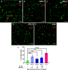
Fig. 3.. In vitro evaluation of hOPC survival after injection with STHs.
(A to E) Representative images of hOPCs after passing through a pulled glass needle (ID = 100 to 200 μm) in the presence of the indicated STH preparations. Con: Control cells were passed through the needle in HBSS, and NS: cells plated without passing through the needle (NS). Scale bars, 200 μm. (F) Quantification of the percentage of dead cells evaluated at 3 hours postseeding using the Live-Dead assay (calcein-ethidium homodimer). Data are presented as mean ± SD. not significant (ns) P ≥ 0.05, *P < 0.05, **P < 0.005, ****P < 0.0001, one-way ANOVA with Dunnett’s post hoc test. n = 3 independent experiments.
Fig. 4.
Fig. 4.. Proliferation of the hOPCs in response to biological cues.
(A and D) Schematic of the experimental setup to evaluate the mitogenic response of hOPCs to HBD-RGD5 peptide or PDGF-AA. The cells were cultured for 48 hours either on (A) wells with the indicated concentrations of immobilized HBD-RGD5 peptide or (D) Lam-coated wells in the presence of the indicated concentrations of PDGF-AA. (B and E) Quantification of the number of hOPCs per square millimeter as a measure of cell growth in response to the (B) HBD-RGD5 peptide or (E) PDGF-AA, respectively. (C and F) Quantification of the hOPC proliferation as percentage of EdU+ cells in response to the biological cues. EdU was detected by labelling with a fluorescently tagged azide via a specific click reaction as per manufacturer’s protocol. (G to I) Representative images of the hOPCs on (G) control/Lam surface, (H) HBD-RGD5 (12.5 nmol/cm2), or (I) PDGF-AA (100 ng/ml). Scale bars, 200 μm. Data are mean ± SD. ns, P ≥ 0.05; *P < 0.05; **P < 0.005; ***P < 0.0005; and ****P < 0.0001; one-way ANOVA with Tukey’s post hoc test. n = 3 independent experiments with three different donors.
Fig. 5.
Fig. 5.. Differentiation of the hOPCs on bidomain fusion peptide, HBD-RGD5.
The in vitro differentiation potential of the hOPCs toward oligodendrocyte fate was evaluated by staining for the oligodendrocyte marker, O4. The cells were cultured on the indicated concentrations of surface immobilized HBD-RGD5 for 4 days in GF-free ND media. (A to F) Representative immunofluorescence images for O4 expression on control/Lam and surfaces with HBD-RGD5 peptide immobilized at the indicated concentrations. Scale bars, 50 μm. (G) Quantification of the number of hOPCs per square millimeter on Lam and HBD-RGD5 immobilized surfaces at the indicated concentrations. (H) Quantification of the percentage of O4+ cells as a function of peptide surface concentration. Data are mean ± SD. ns, P ≥ 0.05; *P < 0.05; **P < 0.005; and ****P < 0.0001; one-way ANOVA with Tukey’s post hoc test. n = 3 independent experiments with three different donors.
Fig. 6.
Fig. 6.. Increased survival of the hOPCs in the NSG mouse brain upon transplantation with STH-2 and immobilized biological cues.
(A) Schematic of the animal transplantation protocol. CD140a+ hOPCs were injected unilaterally into the left hemisphere of the immunocompromised, NSG mice at 100,000/μl within 2 to 3 days of birth. The animals were euthanized at 6 weeks postinjection, and brain sections of 16-μm thickness were collected from rostral to caudal direction throughout the forebrain. The red rectangle indicates the site/point of injection, and the red dots present the transplanted human cells. (B) Number of transplanted hNA+ OPCs in HBSS, STH-2 alone, or STH-2 plus PDGF-AA and HBD-RGD5. Data are mean ± SD. ***P < 0.0005 and ****P < 0.0001, one-way ANOVA with Tukey’s post hoc test. n = 3 animals per condition. (C and D) Representative images of the CC region of the brain sections receiving cells in HBSS or hydrogel. Scale bars, 100 μm. (E) Spatial distribution of hNA+ OPCs injected into the brain CC with either HBSS (control) or STH-2 plus PDGF-AA and HBD-RGD5. Calculation of the area under each curve (AUC) demonstrated 52% higher cell survival in the hydrogel group as compared to control condition. Data are the hNA+ cell count for each brain section per animal, n = 6 to 7 animals per condition, animals were from two different litters, and the transplanted hOPCs were also from with two different donors.
Fig. 7.
Fig. 7.. Increased myelination of the shiverer/rag2 mouse brain upon delivery of the hOPCs with STH-2 fortified with biological signals.
(A and B) Representative confocal microscopy images of the hypomyelinating brain sections immunostained for MBP (green) and hNA (red) at 12 weeks posttransplantation. hOPCs were transplanted either in HBSS (control) or STH-2 + PDGF-AA + HBD-RGD5. (C) Quantification of the myelinated regions (MBP+) of the white matter shows a significant increase in myelination in the STH-2 plus biological cues group compared to the control. Data are mean ± SD. *P < 0.05 and **P < 0.005, two-tailed unpaired t test for the respective brain sections, (D) Quantification of the number of hNA+ OPCs with STH-2 + PDGF-AA + HBD-RGD5 compared to the control. Data are mean ± SD, **P < 0.005, one-tailed unpaired t test. (E to G) Representative sections of mouse brain transplanted with hOPCs in STH-2 + PDGF-AA + HBD-RGD5 stained for markers of oligodendrocytes (OLIG2), postmitotic oligodendrocytes (CC1), and astrocytes (GFAP). White arrowheads indicate staining colocalized with hNA. (H) Quantification of the OLIG2+, CC1+, and GFAP+ cells as a percentage (%) of total hNA+ hOPCs. Data are mean ± SD. ns, P ≥ 0.05, two-tailed unpaired t test between the two groups. (I) Immunofluorescence images of shiverer/rag2 mouse brain stained for MBP (green) and Caspr (red) at 12 weeks posttransplantation. (a) Unmyelinated regions of the brain lacking organized nodes of Ranvier. (b) Densely myelinated region of the CC containing organized nodes of Ranvier, in places where the axonal Caspr protein was in close apposition to human MBP+ internodes. White arrows in (b) indicate normal nodes of Ranvier. n = 4–5 animals per condition from three different litters; transplanted hOPCs were from with three different donors. Scale bars, 500 μm [(A), (B), and (I)], 50 μm [(D) to (F)], and 2.5 μm (a and b).

Similar articles

Cited by

References

    1. Tepavčević V., Lubetzki C., Oligodendrocyte progenitor cell recruitment and remyelination in multiple sclerosis: The more, the merrier? Brain 145, 4178–4192 (2022). - PubMed
    1. Goldman S. A., Mariani J. N., Madsen P. M., Glial progenitor cell-based repair of the dysmyelinated brain: Progression to the clinic. Semin. Cell Dev. Biol. 116, 62–70 (2021). - PMC - PubMed
    1. Windrem M. S., Schanz S. J., Guo M., Tian G. F., Washco V., Stanwood N., Rasband M., Roy N. S., Nedergaard M., Havton L. A., Wang S., Goldman S. A., Neonatal chimerization with human glial progenitor cells can both remyelinate and rescue the otherwise lethally hypomyelinated shiverer mouse. Cell Stem Cell 2, 553–565 (2008). - PMC - PubMed
    1. Sim F. J., McClain C. R., Schanz S. J., Protack T. L., Windrem M. S., Goldman S. A., CD140a identifies a population of highly myelinogenic, migration-competent and efficiently engrafting human oligodendrocyte progenitor cells. Nat. Biotechnol. 29, 934–941 (2011). - PMC - PubMed
    1. Douvaras P., Wang J., Zimmer M., Hanchuk S., O’Bara M. A., Sadiq S., Sim F. J., Goldman J., Fossati V., Efficient generation of myelinating oligodendrocytes from primary progressive multiple sclerosis patients by induced pluripotent stem cells. Stem. Cell Rep. 3, 250–259 (2014). - PMC - PubMed

Publication types

LinkOut - more resources