

Cellular mechanisms of monozygotic twinning: clues from assisted reproduction
- PMID: 38996087
- PMCID: PMC11532623
- DOI: 10.1093/humupd/dmae022
Cellular mechanisms of monozygotic twinning: clues from assisted reproduction
Abstract
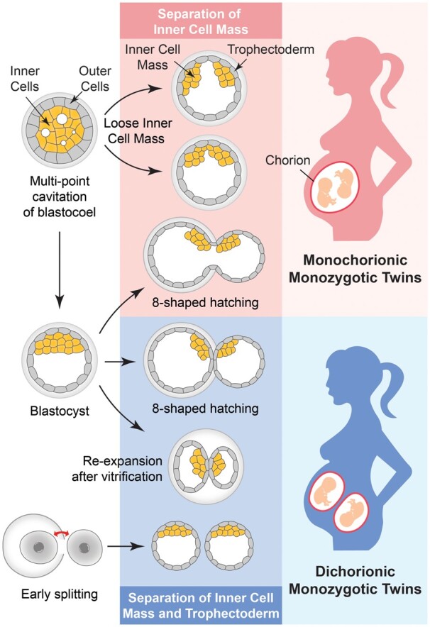
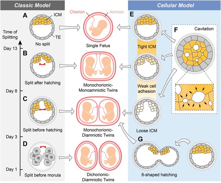
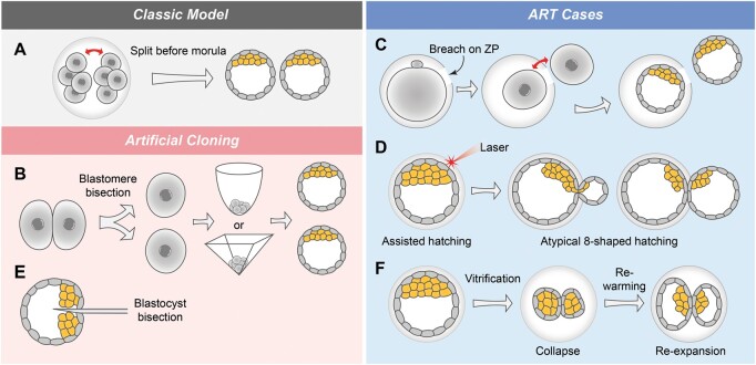
Background: Monozygotic (MZ) twins are believed to arise from the fission of a single fertilized embryo at different stages. Monochorionic MZ twins, who share one chorion, originate from the splitting of the inner cell mass (ICM) within a single blastocyst. In the classic model for dichorionic MZ twins, the embryo splits before compaction, developing into two blastocysts. However, there are a growing number of ART cases where a single blastocyst transfer results in dichorionic MZ twins, indicating that embryo splitting may occur even after blastocyst formation.
Objective and rationale: For monochorionic MZ twins, we conducted a comprehensive analysis of the cellular mechanisms involved in ICM splitting, drawing from both ART cases and animal experiments. In addition, we critically re-examine the classic early splitting model for dichorionic MZ twins. We explore cellular mechanisms leading to two separated blastocysts in ART, potentially causing dichorionic MZ twins.
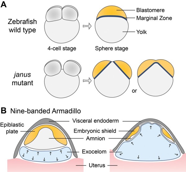
Search methods: Relevant studies including research articles, reviews, and conference papers were searched in the PubMed database. Cases of MZ twins from IVF clinics were found by using combinations of terms including 'monozygotic twins' with 'IVF case report', 'ART', 'single embryo transfer', or 'dichorionic'. The papers retrieved were categorized based on the implicated mechanisms or as those with unexplained mechanisms. Animal experiments relating to MZ twins were found using 'mouse embryo monozygotic twins', 'mouse 8-shaped hatching', 'zebrafish janus mutant', and 'nine-banded armadillo embryo', along with literature collected through day-to-day reading. The search was limited to articles in English, with no restrictions on publication date or species.
Outcomes: For monochorionic MZ twins, ART cases and mouse experiments demonstrate evidence that a looser ICM in blastocysts has an increased chance of ICM separation. Physical forces facilitated by blastocoel formation or 8-shaped hatching are exerted on the ICM, resulting in monochorionic MZ twins. For dichorionic MZ twins, the classic model resembles artificial cloning of mouse embryos in vitro, requiring strictly controlled splitting forces, re-joining prevention, and proper aggregation, which allows the formation of two separate human blastocysts under physiological circumstances. In contrast, ART procedures involving the transfer of a single blastocysts after atypical hatching or vitrified-warmed cycles might lead to blastocyst separation. Differences in morphology, molecular mechanisms, and timing across various animal model systems for MZ twinning can impede this research field. As discussed in future directions, recent developments of innovative in vitro models of human embryos may offer promising avenues for providing fundamental novel insights into the cellular mechanisms of MZ twinning during human embryogenesis.
Wider implications: Twin pregnancies pose high risks to both the fetuses and the mother. While single embryo transfer is commonly employed to prevent dizygotic twin pregnancies in ART, it cannot prevent the occurrence of MZ twins. Drawing from our understanding of the cellular mechanisms underlying monochorionic and dichorionic MZ twinning, along with insights into the genetic mechanisms, could enable improved prediction, prevention, and even intervention strategies during ART procedures.
Registraiton number: N/A.
Keywords: assisted hatching; assisted reproduction; blastocyst cavitation; chorion; embryo development; inner cell mass; monozygotic twins; zona pellucida.
© The Author(s) 2024. Published by Oxford University Press on behalf of European Society of Human Reproduction and Embryology.
Conflict of interest statement
The authors declare that there is no conflict of interest.
Figures
Similar articles
-
Multi-chorionic pregnancies following single embryo transfer at the blastocyst stage: a case series and review of the literature.J Assist Reprod Genet. 2018 Dec;35(12):2109-2117. doi: 10.1007/s10815-018-1329-8. Epub 2018 Oct 25. J Assist Reprod Genet. 2018. PMID: 30362060 Free PMC article. Review.
-
Risk factors associated with monozygotic twinning in offspring conceived by assisted reproductive technology.Hum Reprod Open. 2023 Sep 14;2023(4):hoad035. doi: 10.1093/hropen/hoad035. eCollection 2023. Hum Reprod Open. 2023. PMID: 37840637 Free PMC article.
-
First case of dichorionic diamniotic triplet pregnancy after single blastocyst transfer.J Assist Reprod Genet. 2024 Feb;41(2):437-440. doi: 10.1007/s10815-023-02989-4. Epub 2023 Dec 11. J Assist Reprod Genet. 2024. PMID: 38079075 Free PMC article.
-
Determinants of monozygotic twinning in ART: a systematic review and a meta-analysis.Hum Reprod Update. 2018 Jul 1;24(4):468-483. doi: 10.1093/humupd/dmy006. Hum Reprod Update. 2018. PMID: 29538675
-
Developmental clock compromises human twin model created by embryo splitting.Hum Reprod. 2015 Dec;30(12):2774-84. doi: 10.1093/humrep/dev252. Epub 2015 Oct 21. Hum Reprod. 2015. PMID: 26489438
Cited by
-
Development of external morphological malformations induced by hyperthermia exposure during the blastula stage in an ex-ovo (shell-less) culture of Gallus gallus domesticus embryos.Poult Sci. 2025 Aug;104(8):105341. doi: 10.1016/j.psj.2025.105341. Epub 2025 May 26. Poult Sci. 2025. PMID: 40446690 Free PMC article.
-
Examining the Utility of the Mammalian Methylation Array for Pan-Mammalian Analysis of Monozygotic Twinning.Epigenomes. 2024 Oct 6;8(4):37. doi: 10.3390/epigenomes8040037. Epigenomes. 2024. PMID: 39449361 Free PMC article.
References
-
- Abdelilah S, Driever W.. Pattern formation in janus-mutant zebrafish embryos. Dev Biol 1997;184:70–84. - PubMed
-
- Abdelilah S, Solnica-Krezel L, Stainier DY, Driever W.. Implications for dorsoventral axis determination from the zebrafish mutation janus. Nature 1994;370:468–471. - PubMed
-
- Alikani M, Cekleniak NA, Walters E, Cohen J.. Monozygotic twinning following assisted conception: an analysis of 81 consecutive cases. Hum Reprod 2003;18:1937–1943. - PubMed
-
- Alikani M, Noyes N, Cohen J, Rosenwaks Z.. Monozygotic twinning in the human is associated with the zona pellucida architecture. Hum Reprod 1994;9:1318–1321. - PubMed
Publication types
MeSH terms
Grants and funding
LinkOut - more resources
Full Text Sources
Medical
Research Materials
