Review
. 2024 Sep;11(5):e200275.
doi: 10.1212/NXI.0000000000200275. Epub 2024 Jul 12.

MOG CNS Autoimmunity and MOGAD

Affiliations
Review

MOG CNS Autoimmunity and MOGAD

Carson E Moseley et al. Neurol Neuroimmunol Neuroinflamm. 2024 Sep.

Abstract

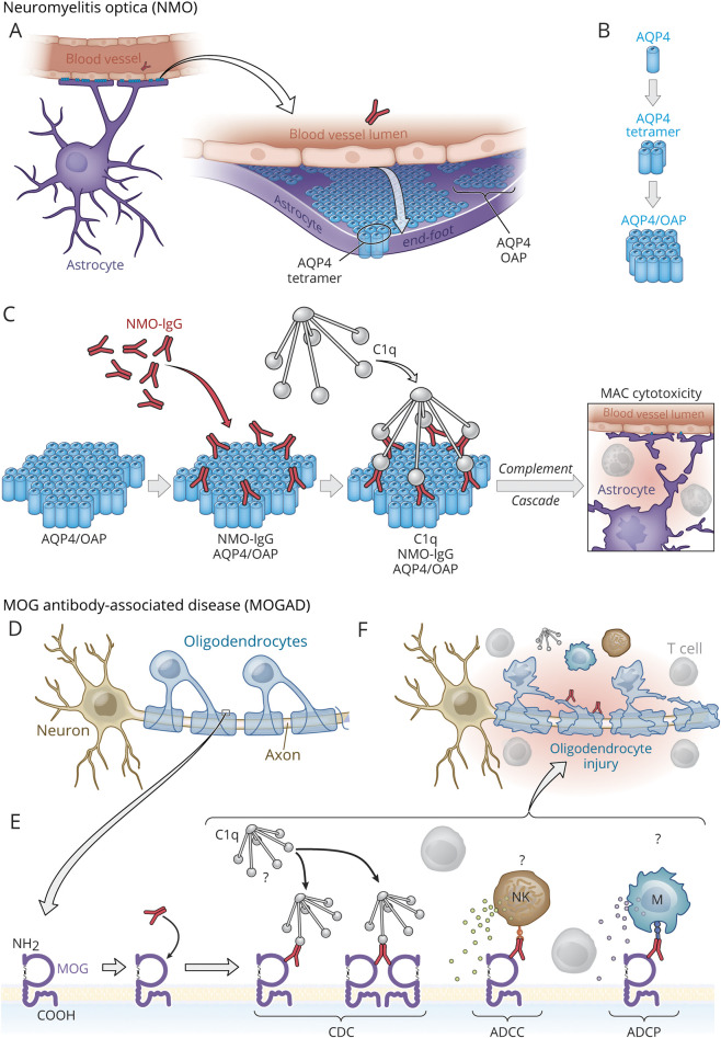
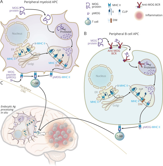
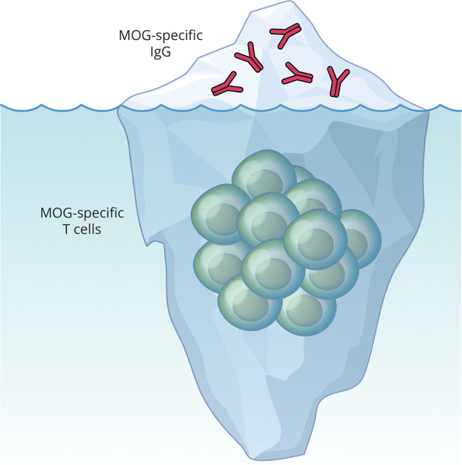
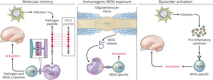
At one time considered a possible form of neuromyelitis optica (NMO) spectrum disorder (NMOSD), it is now accepted that myelin oligodendrocyte glycoprotein (MOG) antibody (Ab)-associated disorder (MOGAD) is a distinct entity from either NMO or multiple sclerosis (MS) and represents a broad spectrum of clinical phenotypes. Whereas Abs targeting aquaporin-4 (AQP4) in NMO are pathogenic, the extent that anti-MOG Abs contribute to CNS damage in MOGAD is unclear. Both AQP4-specific Abs in NMO and MOG-specific Abs in MOGAD are predominantly IgG1, a T cell-dependent immunoglobulin (Ig) subclass. Key insights in neuroimmunology and MOGAD pathogenesis have been learned from MOG experimental autoimmune encephalomyelitis (EAE), described 2 decades before the term MOGAD was introduced. MOG-specific T cells are required in MOG EAE, and while anti-MOG Abs can exacerbate EAE and CNS demyelination, those Abs are neither necessary nor sufficient to cause EAE. Knowledge regarding the spectrum of MOGAD clinical and radiologic presentations is advancing rapidly, yet our grasp of MOGAD pathogenesis is incomplete. Understanding both the humoral and cellular immunology of MOGAD has implications for diagnosis, treatment, and prognosis.


Conflict of interest statement

The authors report no relevant disclosures. Go to Neurology.org/NN for full disclosures.

Figures

Figure 1
Figure 1. Models Illustrating How Antibodies and Complement May Participate in NMO and MOGAD
AQP4, a transmembrane (TM) protein, is expressed in the astrocytes, kidneys, muscle, and lung, yet the AQP4-specific IgG1 in NMO damages astrocytes primarily., AQP4 is expressed abundantly in astrocyte end-foot processes at the blood-brain barrier (BBB) (A), where tetramers of AQP4 (M23:M23 and M23:M21 isoforms) assemble into orthogonal arrays of particles (OAPs) (B), which provides a lattice-like platform that permits monovalent binding of AQP4-specific IgG1 in a distribution that is optimal for recruitment of C1q (C) and initiation of the classical complement cascade that results in cytotoxicity to astrocytes from formation of the membrane attack complex (MAC). (D–G) MOG, also a TM protein, is exposed on the outer oligodendrocyte surface. Unlike AQP4-specific IgG1 in NMO, the role of anti-MOG antibodies in CNS damage and the potential mechanisms (e.g., complement-dependent cytotoxicity [CDC], antibody-dependent cellular cytotoxicity [ADCC], or antibody-dependent cellular phagocytosis [ADCP]) employed by MOG-specific IgG1 in MOGAD are less clear. Complement deposition is identified inconsistently in MOGAD lesions. Unlike AQP4-specific IgG1 from patients with NMO, MOG-specific IgG1 from patients with MOGAD may preferentially bind MOG in a bivalent manner that is less efficient for recruitment of C1q and activation of CDC but may promote ADCC or ADCP. Natural killer cell (NK); myeloid APC (M). Copyright Xavier Studio, reprinted with permission.
Figure 2
Figure 2. MHC II Endocytic Antigen Processing and Presentation to T Cells in MOG CNS Autoimmunity
This figure illustrates types of APCs, including myeloid cells (A), B cells (B), and microglia (C) that may participate in MOG CNS autoimmunity. MOG EAE is typically induced by subcutaneous immunization with either MOG protein or MOG peptide (pMOG), which leads to peripheral (outside CNS) activation of pathogenic MOG-specific T cells that traffic into the CNS and initiate CNS inflammation. Human MOG protein, which contains proline-42, causes T-B-dependent EAE., MOG-specific T-cell activation requires recognition of pMOG in association with MHC II molecules expressed on APCs. Antigen processing of MOG protein through the MHC II endocytic pathway is required in at least at 2 stages in MOG protein-induced EAE, for initial recognition by MOG-specific T cells in the periphery and for reactivation within the CNS (in situ)., Several molecules, including invariant chain (Ii, CD74), HLA-DM (H-2M) (DM), and proteases participate in MHC II maturation and in orchestrating steps within the endocytic pathway. MHC II (α/β) molecules associate with Ii in the endoplasmic reticulum (ER) forming a trimer (Ii-MHC II) and travel through the Golgi to the endosomal compartment. Ii is enzymatically degraded yielding a fragment of Ii, class II Ii peptide (CLIP), which remains bound within the MHC II peptide-binding groove. The MHC II chaperone DM facilitates removal of CLIP, permitting exchange for antigenic peptide (e.g., pMOG). Peripheral myeloid cells capture native antigenic proteins (MOG) (top of A) via phagocytosis or pinocytosis and deliver them to the endolysosomal compartment where they are degraded by proteases into 9–14 amino acid fragments that can bind the MHC II peptide-binding groove. Endosomes containing peptide-MHC II complexes fuse with the plasma membrane permitting presentation of peptides (e.g., pMOG-MHC II) to encephalitogenic MOG-specific T cells. In contrast to MOG protein, pMOG immunization (lower left in A) supplies peptide that can bind cell surface MHC II molecules directly, bypassing the need for endocytic processing. B cells are exceptionally efficient APCs when they bind antigens (e.g., MOG protein) (top of B) with their B-cell antigen-specific receptors (anti-MOG-BCR) and deliver them to the endolysosomal compartment for processing and association with MHC II molecules., Independent of whether MOG EAE is induced by immunization with MOG protein, pMOG, or by adoptive transfer of encephalitogenic MOG-specific T cells (not shown), endocytic Ag processing by APCs (e.g., resident microglia) (shown in C) in situ is required for recognition of cognate MOG peptide by MOG-specific T cells that initiate CNS inflammation., Copyright Xavier Studio, reprinted with permission.
Figure 3
Figure 3. Cellular Immunity May Have a Prominent Role in MOGAD Pathogenesis
MOG IgG (above water surface) is necessary for diagnosis of MOGAD, yet cellular immunity (beneath water surface) is required in models of MOG CNS autoimmunity. MOG-specific antibodies in MOGAD and MOG EAE are IgG, a T-cell-dependent Ig subclass. CD4+ T cells predominate in MOGAD lesions and in EAE lesions. Other cells (e.g., resident microglia, infiltrating myeloid cells, fewer CD8+ T cells, and B cells) also contribute to CNS inflammation (not depicted). In general, MOG-specific antibodies do not induce disease in the absence of inflammation. MOG-specific T cells can induce EAE in the absence of B cells or antibodies., Collectively, such findings suggest that seronegative MOG-targeted autoimmune disease (“sMOG”) may exist in some individuals who manifest clinical symptoms associated with MOGAD in the absence of detectable MOG-specific antibodies. Graphics created using BioRender.com. Copyright Xavier Studio, reprinted with permission.
Figure 4
Figure 4. Potential Triggers of MOG Autoimmunity
Several possible mechanisms may promote MOG CNS autoimmune disease. MOG autoimmunity could be triggered by molecular mimicry (left panel), a process that can occur when antigenic determinants of pathogens cross-react with self-antigens. Immunogenic MOG exposure (middle panel), either from CNS damage secondary from another disease, or ectopic MOG expression could promote activation of MOG-specific immune cells. The normal immune repertoire contains MOG-reactive T cells. Thus, bystander activation (right panel) of preexisting MOG-specific T cells by proinflammatory cytokines from an unrelated stimulus (e.g., systemic infection) could theoretically lead to proinflammatory polarization and expansion of MOG-specific T cells. Copyright Xavier Studio, reprinted with permission.

References

    1. Lennon VA, Kryzer TJ, Pittock SJ, Verkman AS, Hinson SR. IgG marker of optic-spinal multiple sclerosis binds to the aquaporin-4 water channel. J Exp Med. 2005;202(4):473-477. doi:10.1084/jem.20050304 - DOI - PMC - PubMed
    1. Linnington C, Webb M, Woodhams PL. A novel myelin-associated glycoprotein defined by a mouse monoclonal antibody. J Neuroimmunol. 1984;6:387-396. doi:10.1016/0165-5728(84)90064-x - DOI - PubMed
    1. Kerlero de Rosbo N, Mendel I, Ben-Nun A. Chronic relapsing experimental autoimmune encephalomyelitis with a delayed onset and an atypical clinical course, induced in PL/J mice by myelin oligodendrocyte glycoprotein (MOG)-derived peptide: preliminary analysis of MOG T cell epitopes. Eur J Immunol. 1995;25(4):985-993. doi:10.1002/eji.1830250419 - DOI - PubMed
    1. Kerlero de Rosbo N, Milo R, Lees MB, Burger D, Bernard CC, Ben-Nun A. Reactivity to myelin antigens in multiple sclerosis. Peripheral blood lymphocytes respond predominantly to myelin oligodendrocyte glycoprotein. J Clin Invest. 1993;92(6):2602-2608. doi:10.1172/jci116875 - DOI - PMC - PubMed
    1. Berger T, Rubner P, Schautzer F, et al. . Antimyelin antibodies as a predictor of clinically definite multiple sclerosis after a first demyelinating event. N Engl J Med. 2003;349(2):139-145. doi:10.1056/nejmoa022328 - DOI - PubMed

MeSH terms

LinkOut - more resources