. 2024 Sep:199:106590.
doi: 10.1016/j.nbd.2024.106590. Epub 2024 Jul 10.

Withdrawal from chronic alcohol impairs the serotonin-mediated modulation of GABAergic transmission in the infralimbic cortex in male rats

Affiliations

Withdrawal from chronic alcohol impairs the serotonin-mediated modulation of GABAergic transmission in the infralimbic cortex in male rats

Roman Vlkolinsky et al. Neurobiol Dis. 2024 Sep.

Abstract

The infralimbic cortex (IL) is part of the medial prefrontal cortex (mPFC), exerting top-down control over structures that are critically involved in the development of alcohol use disorder (AUD). Activity of the IL is tightly controlled by γ-aminobutyric acid (GABA) transmission, which is susceptible to chronic alcohol exposure and withdrawal. This inhibitory control is regulated by various neuromodulators, including 5-hydroxytryptamine (5-HT; serotonin). We used chronic intermittent ethanol vapor inhalation exposure, a model of AUD, in male Sprague-Dawley rats to induce alcohol dependence (Dep) followed by protracted withdrawal (WD; 2 weeks) and performed ex vivo electrophysiology using whole-cell patch clamp to study GABAergic transmission in layer V of IL pyramidal neurons. We found that WD increased frequencies of spontaneous inhibitory postsynaptic currents (sIPSCs), whereas miniature IPSCs (mIPSCs; recorded in the presence of tetrodotoxin) were unaffected by either Dep or WD. The application of 5-HT (50 μM) increased sIPSC frequencies and amplitudes in naive and Dep rats but reduced sIPSC frequencies in WD rats. Additionally, 5-HT2A receptor antagonist M100907 and 5-HT2C receptor antagonist SB242084 reduced basal GABA release in all groups to a similar extent. The blockage of either 5-HT2A or 5-HT2C receptors in WD rats restored the impaired response to 5-HT, which then resembled responses in naive rats. Our findings expand our understanding of synaptic inhibition in the IL in AUD, indicating that antagonism of 5-HT2A and 5-HT2C receptors may restore GABAergic control over IL pyramidal neurons. SIGNIFICANCE STATEMENT: Impairment in the serotonergic modulation of GABAergic inhibition in the medial prefrontal cortex contributes to alcohol use disorder (AUD). We used a well-established rat model of AUD and ex vivo whole-cell patch-clamp electrophysiology to characterize the serotonin modulation of GABAergic transmission in layer V infralimbic (IL) pyramidal neurons in ethanol-naive, ethanol-dependent (Dep), and ethanol-withdrawn (WD) male rats. We found increased basal inhibition following WD from chronic alcohol and altered serotonin modulation. Exogenous serotonin enhanced GABAergic transmission in naive and Dep rats but reduced it in WD rats. 5-HT2A and 5-HT2C receptor blockage in WD rats restored the typical serotonin-mediated enhancement of GABAergic inhibition. Our findings expand our understanding of synaptic inhibition in the infralimbic neurons in AUD.

Keywords: Alcohol use disorder; Electrophysiology; GABA; Infralimbic cortex; Patch-clamp; Serotonin.


Conflict of interest statement

Declaration of competing interest The authors declare no competing financial interests.

Figures
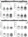
Fig. 1.
Fig. 1.
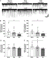
Extended alcohol withdrawal increases basal GABA transmission in IL pyramidal neurons without effect on AP-independent, stochastic GABA release. (A) Representative traces of sIPSCs in naive, Dep, and WD neurons. (B) Baseline frequency of the sIPSCs in WD neurons was significantly higher than frequencies in the naive or Dep groups (Tukey’s post hoc test, *p < 0.05, **p < 0.01). (C) sIPSC amplitudes were comparable across groups. (D) sIPSC rise times were significantly prolonged in the WD group compared with the naive group (Tukey’s post hoc test, *p < 0.05), but (E) decay times remained comparable across all groups. (F) Representative traces of mIPSCs in naive, Dep, and WD animals recorded in IL layer V pyramidal neurons in the presence of 0.5 μM TTX. (G) mIPSC frequency representing AP-independent presynaptic, stochastic GABA release was unaffected by alcohol exposure or protracted WD. (H-J) Postsynaptic mIPSC parameters were unaffected by alcohol exposure or protracted WD. The data are expressed as the mean ± SEM.
Fig. 2.
Fig. 2.
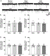
Serotonin-mediated enhancement of GABAergic inhibition in IL layer V pyramidal neurons is impaired in WD rats. (A) Representative traces of sIPSCs in naive, Dep, and WD neurons. (B) In the naive and Dep groups, exogenously applied 5-HT significantly increased sIPSC frequencies (#p < 0.05) compared with their baseline values (dashed line, 100%). In the WD group, we observed a reversal of this response and overall decreases in mean sIPSC frequencies (#p < 0.05), leading to significant differences between the naive and WD groups (Tukey’s post hoc test, *p < 0.05). (C) Serotonin significantly increased sIPSC amplitudes in the naive and Dep groups (#p < 0.05) compared with their baseline values (dashed line, 100%), but this enhancing effect was blunted in the WD group, leading to a significant difference between the naive and WD groups (Tukey’s post hoc test, *p < 0.05). (D) Serotonin application significantly increased the sIPSC rise time in the Dep group only (#p < 0.05). (E) sIPSC decay times were prolonged in the naive and Dep groups compared to their baselines (dashed line, 100%; #p < 0.05), and such prolongation was absent in the WD group, leading to a significant difference between the naive and WD groups (Tukey’s post hoc test, *p < 0.05). The altered response to 5-HT indicates neuroadaptations that are induced by alcohol dependence and/or protracted WD. The data are expressed as the mean ± SEM.
Fig. 3.
Fig. 3.
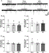
Serotonin does not enhance AP-independent, stochastic presynaptic GABA release in IL pyramidal neurons in naive, Dep, or WD male rats. (A) Representative traces of mIPSCs recorded in the presence of 0.5 μM TTX in neurons from naive, Dep, and WD rats. (B-E) Frequency, amplitudes, and rise and decay times were not significantly affected by exogenously applied 5-HT. The data are expressed as the mean ± SEM.
Fig. 4.
Fig. 4.
5-HT2A receptor antagonist M100907 inhibits basal sIPSC frequencies in IL pyramidal neurons in all groups. (A) Representative sIPSCs in IL neurons from naive, Dep, and WD rats. (B) M100907 application (100 nM) reduced sIPSC frequencies compared to their baselines (dashed line, 100%) in all groups to a similar extent, indicating lower presynaptic GABA release (one-sample t-test, #p < 0.05 and ##p < 0.01). (C) sIPSCs amplitudes were unaffected by the antagonist but in the naive group only M100907 prolonged sIPSC rise (D) and decay (E) times compared to their baselines (dashed line, 100%; one-sample t-test, #p < 0.05). The data are expressed as the mean ± SEM.
Fig. 5.
Fig. 5.
Interaction between 5-HT2A receptor antagonist M100907 and exogenous 5-HT in naive, Dep, and WD rats. (A) Representative sIPSCs from pyramidal neurons perfused with M100907 (100 nM, left) followed by co-application of 5-HT (50 μM, right). (B). In all groups, 5-HT application in the presence of M100907 significantly increased the mean sIPSC frequency when compared to the last time bin of antagonist application (dashed line, 100%, one-sample t-test, #p < 0.05, ##p < 0.01). (C) Pair-wise comparison of sIPSC frequencies in individual pyramidal neurons between M100907 alone and 5-HT co-applied with M100907. M100907 reduced the mean sIPSC frequency in all groups. In the naive group, 5-HT co-application with M100907 enhanced sIPSC frequency in seven neurons but reduced it in the remaining 3 neurons, with an overall significant increasing effect. Similarly, in the presence of M100907, 5-HT increased sIPSC frequencies in seven of eight neurons in Dep neurons, and in all WD neurons. RM-ANOVA followed by Tukey’s multiple-comparison post hoc test confirmed significant increases in frequencies in all groups. (Tukey’s *p < 0.05, ***p < 0.001). Scatter plots indicate values in individual neurons. Bars represent means. (D–F) Postsynaptic measures, including sIPSC amplitude and rise and decay times, were unaffected by 5-HT application in any group. The data are expressed as the mean ± SEM.
Fig. 6.
Fig. 6.
5-HT2C receptor antagonist SB242084 decreases basal GABA transmission in all groups. (A) Representative sIPSCs were recorded in IL neurons from naive, Dep, and WD rats. (B) The application of SB242084 (100 nM) significantly decreased sIPSC frequencies compared to their baseline (dashed line, 100%) in the naive, Dep, and WD groups (one sample t-test, #p < 0.05, ##p < 0.05). Between-group comparisons did not identify statistically significant differences indicating similar size of inhibition in all groups (one-way ANOVA). (C-E) Postsynaptic parameters (amplitude, rise and decay times) were not significantly changed by SB242084 application. The data are expressed as the mean ± SEM.
Fig. 7.
Fig. 7.
Interaction of 5-HT2C receptor antagonist SB242084 and exogenous 5-HT in naive, Dep, and WD rats. (A) Representative sIPSCs from pyramidal neurons perfused with SB242084 (100 nM, left) followed with co-application of 5-HT (50 μM, right). (B) In all groups, 5-HT application in the presence of SB242084 significantly increased the mean sIPSC frequency when compared to the last time bin of antagonist application (dashed line, 100%, one-sample t-test, #p < 0.05, ##p < 0.01). Notably, the 5-HT-induced increases in sIPSC frequencies in the WD group exceeded increases in the naive and Dep groups (one-way ANOVA, p = 0.02; Tukey’s post hoc test, *p < 0.05, vs. naïve; *p = 0.02, vs. Dep). The data are expressed as the mean ± SEM. (C) Pair-wise comparison of sIPSC frequencies in individual pyramidal neurons between SB242084 alone and 5-HT co-applied with SB242084. SB242084 reduced the mean sIPSC frequency but subsequent 5-HT co-application consistently enhanced frequencies in all neurons in all experimental groups. RM-ANOVA followed by Tukey’s post hoc confirmed statistically significant enhancement in the naïve, Dep, and WD groups (Tukey’s *p < 0.05, ***p < 0.001). Scatter plots indicate relative values in individual neurons. Bars represent means. (D–F) The presence of SB242084 blocked effects of 5-HT on postsynaptic sIPSC parameters in the naive and Dep groups but significantly increased amplitudes in the WD group (one-sample t-test, #p < 0.05). Thus, there were significant differences between the WD group vs. naive and Dep groups (Tukey’s post hoc test following one-way ANOVA, **p < 0.01). sIPSC rise times were not significantly affected by 5-HT in any group, while decay times were significantly prolonged in the WD group only (one-sample t-test, #p < 0.05). The data are expressed as the mean ± SEM.
Fig. 8.
Fig. 8.
Effects of chronic-intermittent alcohol exposure on passive neuronal membrane properties in IL layer V pyramidal neurons. (A) Current to voltage relationship (I-V) recorded in current-clamp mode. To determine Vm, linear fits were constructed in each group at a range from −120 to +100 pA (before AP generation) at 15 pA increments. For clarity, 30 pA steps are indicated. Mean slopes of I-V fits in naive (open circles), Dep (gray), and WD (black) rats did not significantly differ between groups, indicating no change in passive membrane properties. (Inset) Representative voltage traces in response to incremental current (30 pA) steps in pyramidal neurons in the naive group. (B) Linear current vs. Vmsag relationship. Withdrawal reduced the maximal Vmsag (Max Vmsag) determined and compared at −120 pA (One way ANOVA, also see Tables S2–3). Representative traces of incremental hyperpolarizing current steps at a range of −120 to 0 pA in naive pyramidal neurons (bottom traces). The upper traces represent voltage responses and measurement of the maximal voltage sag. The data are expressed as the mean ± SEM.

References

    1. Abernathy K., et al. , 2010. Alcohol and the prefrontal cortex. Int. Rev. Neurobiol 91, 289–320. - PMC - PubMed
    1. Abi-Saab WM, et al. , 1999. 5-HT2 receptor regulation of extracellular GABA levels in the prefrontal cortex. Neuropsychopharmacology 20, 92–96. - PubMed
    1. Arvanov VL, Wang RY, 1998. M100907, a selective 5-HT2A receptor antagonist and a potential antipsychotic drug, facilitates N-methyl-D-aspartate-receptor mediated neurotransmission in the rat medial prefrontal cortical neurons in vitro. Neuropsychopharmacology 18, 197–209. - PubMed
    1. Athilingam JC, et al. , 2017. Serotonin enhances excitability and gamma frequency temporal integration in mouse prefrontal fast-spiking interneurons. Elife 6. - PMC - PubMed
    1. Borgonetti V., et al. , 2023. IL-18 signaling in the rat central amygdala is disrupted in a comorbid model of post-traumatic stress and alcohol use disorder. Cells 12. - PMC - PubMed

Publication types

MeSH terms