. 2024 Aug;47(8):100091.
doi: 10.1016/j.mocell.2024.100091. Epub 2024 Jul 10.

Decrease of alpha-crystallin A by miR-325-3p in retinal cells under blue light exposure

Affiliations

Decrease of alpha-crystallin A by miR-325-3p in retinal cells under blue light exposure

Subeen Oh et al. Mol Cells. 2024 Aug.

Abstract

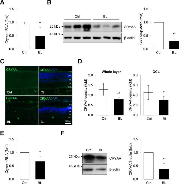
Exposure to blue light can lead to retinal degeneration, causing adverse effects on eye health. Although the loss of retinal cells due to blue light exposure has been observed, the precise molecular mechanisms underlying this process remain poorly understood. In this study, we investigate the role of alpha-crystallin A (CRYAA) in neuro-retinal degeneration and their regulation by blue light. We observed significant apoptotic cell death in both the retina of rats and the cultured neuro-retinal cells. The expressions of Cryaa mRNA and protein were significantly downregulated in the retina exposed to blue light. We identified that miR-325-3p reduces Cryaa mRNA and protein by binding to its 3'-untranslated region. Upregulation of miR-325-3p destabilized Cryaa mRNA and suppresses CRYAA, whereas downregulation of miR-325-3p increased both expressions. Blue light-induced neuro-retinal cell death was alleviated by CRYAA overexpression. These results highlight the critical role of Cryaa mRNA and miR-325-3p molecular axis in blue light-induced retinal degeneration. Consequently, targeting CRYAA and miR-325-3p presents a potential strategy for protecting against blue light-induced retinal degeneration.

Keywords: Alpha-crystallin A; High-energy visible light; MicroRNAs; Neuro-retinal cell; Retina.


Conflict of interest statement

Declaration of Competing Interests The authors declare that they have no known competing financial interests or personal relationships that could have appeared to influence the work reported in this paper.

Figures

Fig. 1
Fig. 1
Blue light induces cell death in neuro-retinal cells. (A) Rat and R-28 cells were exposed with or without blue light (460 nm) in a chamber set at 2,000 lux for 120 minutes and 10 minutes, respectively. The sacrifice was performed 3 days after the blue light exposure. (B, C) Representative images of retina tissues and R-28 cells stained with TUNEL (Red) and DAPI (blue) after blue-light irradiation. TUNEL-positive cells are observed throughout the entire retinal layer, including the GCL. Scale bars represent 50 µm and 100 µm. (D, E) Quantitative analysis for the number of TUNEL-positive cells. (E) The viability of R-28 cells exposed to blue light or not was determined by MTT assay. **P < .01; ***P < .001. DAPI, 4′,6-diamidino-2-phenylindole; GCL, ganglion cell layer.
Fig. 2
Fig. 2
Exposure to blue light caused reducing the expression of Cryaa mRNA and protein. (A, B) After blue light exposure, levels of Cryaa mRNA and protein in the retinal tissues of rats were analyzed by RT-qPCR and western blotting. (C) Representative immunoreactivity of CRYAA in the retinal tissues of rats exposed to blue light. CRYAA (green) is expressed in the GCL and OS surrounding the nuclei of neuro-retinal cells (DAPI: nuclear staining, blue). (D) A quantitative analysis of the density of CRYAA is shown. (E, F) levels of Cryaa mRNA and protein in blue light exposure R-28 cells were determined by RT-qPCR and western blotting. Gapdh mRNA was used for normalization, and β-actin was used as a loading control. Data represent the means ± SEM from 3 independent experiments. *P < .05; **P < .01. DAPI, 4′,6-diamidino-2-phenylindole; GCL, ganglion cell layer; OS, outer segments.
Fig. 3
Fig. 3
The miR-325-3p regulates Cryaa mRNA and protein expressions in neuro-retinal cells. (A, B) After blue-light exposure, miR-325-3p levels in the retinal tissues of rats and R-28 cells were quantified by RT-qPCR. (C) After transfection of R-28 cells with miR-325-3p mimic or inhibitor and appropriate control (miR-Ctrl), levels of miR-325-3p were quantified by RT-qPCR. Levels of Cryaa mRNAs (D, F) and CRYAA proteins (E, G) in R-28 cells were analyzed by RT-qPCR and western blotting, respectively. U6 RNA, and Gapdh mRNA were used for normalization, and β-actin was used as a loading control. Data represent the means ± SEM from 3 independent experiments. *P < .05; **P < .01.
Fig. 4
Fig. 4
MiR-325-3p regulates Cryaa mRNA by targeting its 3′UTR. (A) Schematic of reporter constructs. 3′UTR of Cryaa mRNA containing a miR-325-3p binding site was inserted into pEGFP-C1. A mutant reporter construct lacking the miR-325-3p binding site was generated using site-directed mutagenesis. (B) After transfection to R-28 cells with miR-325-3p mimic, miR-325-3p inhibitor, and appropriate control (miR-Ctrl), together with each reporter plasmid, levels of GFP were assessed by western blotting. β-actin was used as a loading control. Data represent the means ± SEM from 3 independent experiments.
Fig. 5
Fig. 5
Blue light reduces neuro-retinal cell viability through Cryaa mRNA and miR-325-3p axis. (A) After transfection with siCryaa and appropriate control (siCtrl), the viability of R-28 cells exposed with or without blue light was determined by MTT assay. (B) After transfection with plasmid pCMV-Cryaa (pCryaa) and appropriate control (pCtrl), the viability of R-28 cells exposed with or without blue light was determined by MTT assay. (C, D) Representative levels of CRYAA protein in R-28 cells (transfected with siRNA or plasmid) were determined by western blotting. β-actin was used as a loading control. Data represent the means ± SEM from 3 independent experiments. *P < .05.

References

    1. Aggio-Bruce R., Chu-Tan J.A., Wooff Y., Cioanca A.V., Schumann U., Natoli R. Inhibition of microRNA-155 protects retinal function through attenuation of inflammation in retinal degeneration. Mol. Neurobiol. 2021;58:835–854. - PMC - PubMed
    1. Aiello L.P. Suppression of retinal neovascularization in vivo by inhibition of vascular endothelial growth factor (VEGF) using soluble VEGF-receptor chimeric proteins. Proc. Natl. Acad. Sci. U.S.A. 1995;92:10457–10461. - PMC - PubMed
    1. Andley U.P. Crystallins in the eye: function and pathology. Prog. Retin. Eye Res. 2007;26:78–98. - PubMed
    1. Arac A., Brownell S.E., Rothbard J.B., Chen C., Ko R.M., Pereira M.P., Albers G.W., Steinman L., Steinberg G.K. Systemic augmentation of alphaB-crystallin provides therapeutic benefit twelve hours post-stroke onset via immune modulation. Proc. Natl. Acad. Sci. U.S.A. 2011;108:13287–13292. - PMC - PubMed
    1. Athanasiou D., Aguila M., Bevilacqua D., Novoselov S.S., Parfitt D.A., Cheetham M.E. The cell stress machinery and retinal degeneration. FEBS Lett. 2013;587:2008–2017. - PMC - PubMed

LinkOut - more resources