. 2024 Jul 12;10(1):322.
doi: 10.1038/s41420-024-02080-6.

PTBP1 protects Y RNA from cleavage leading to its apoptosis-specific degradation

Affiliations

PTBP1 protects Y RNA from cleavage leading to its apoptosis-specific degradation

Takeshi Kamakura et al. Cell Death Discov. .

Abstract

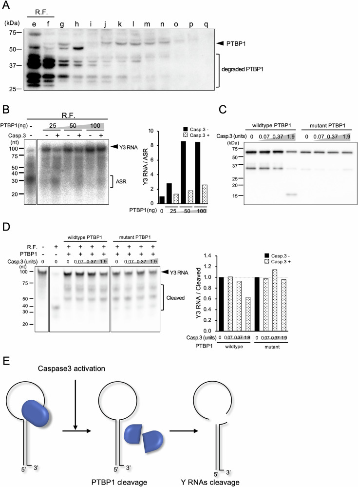
Some RNAs such as 28S rRNA, U1 small nuclear RNA (snRNA), and Y RNAs are known to be cleaved during apoptosis. The underlying mechanism, functions, and biological significance of RNA degradation in apoptosis remain elusive. Y RNAs are non-coding RNAs widely conserved from bacteria to mammals, and are major components of Ro ribonucleoprotein (RNP) complexes which contain the 60 kDa Ro protein (SS-A) and the 50 kDa La protein (SS-B). The autoantigenic Ro and La proteins were identified by autoantibodies present in the sera from patients with Systemic lupus erythematosus (SLE) and Sjögren's syndrome (SjS). We previously identified novel, functional small RNAs named AGO-taxis small RNAs (ASRs) that are specifically bound to Argonaute protein 1 (AGO1), which are processed from Y RNAs. Cell-free analysis combined with fractionation methods revealed that the apoptosis-specific biogenesis of ASRs or cleavage of Y RNA was induced by truncation of polypyrimidine tract-binding protein 1 (PTBP1), which is an endoribonuclease inhibitor of Y RNAs by caspase 3. Caspase 3-resistant PTBP1 mutant protected cleavage of Y RNAs in apoptosis induced by staurosporine. Furthermore, caspase 3-resistant PTBP1 mutant knock-in mice showed elevated cytokines, dysregulation of the germinal center formation compared to the wild-type mice at LPS stimulation, and high positivity of antinuclear antibody. Those results suggest that cleavage of Y RNAs or biogenesis of ASR during apoptosis has critical biological functions and their deregulation result in immune dysregulation and the formation of autoantibody, possibly leading to the development of autoimmune diseases.


Conflict of interest statement

The authors declare no competing interests.

Figures

Fig. 1
Fig. 1. Y RNA cleavage is activated in apoptotic cells.
A Common motif of ASRs was identified by MEME motif search. The motif, indicated in the capital letter, was plotted on Y RNA common structure. Sequences of the most abundant ASRs from each Y RNA were analyzed. B EBV lytic infection was induced by BCR stimulation using anti-human IgG antibody. After 24 h, apoptosis induction was investigated by flow cytometry. Annexin V single positivity shows early apoptotic cells and double positivity shows late apoptotic cells. C Y3 RNA and ASRs cleavage product were detected by northern blotting for latent or lytic infection of EBV (C), and with or without caspase 3 (D). Similar results were obtained in two independent experiments.
Fig. 2
Fig. 2. Y RNA processing is independent of DROSHA, DICER, and RNase L.
A Expression of DROSHA and DICER in the Jurkat cells transfected with control siRNA (Ctrl.), siDROSHA, or siDICER by real-time PCR (n = 3) normalized by GAPDH (Left). Expression of Y RNA fragments in those cells, where apoptosis was induced by anti-Fas antibody (clone: CH-11), by northern blotting (Right). Upper panel, Y3 RNA and ASR; lower panel, Y5 RNA and ASR. B Dicer-knockout or wild-type (floxed Dicer) MEFs were cultivated with or without 10 µM staurosporine for 2 h, followed by the detection of Y3 RNA cleavage by northern blotting. C Expression of RNase L in the Jurkat cells transfected with control siRNA (Ctrl.) or siRNase L by real-time PCR (n = 3) normalized by GAPDH. Y3 RNA or Y5 RNA cleavage was also detected by northern blotting as in (A). D Rnasel-knockout or wild-type MEFs were stimulated with 10 μM staurosporine for 2 or 6 h, followed by the detection of Y3 RNA cleavage by northern blotting. Similar results were obtained in two independent experiments. Student’s t-test was used for the statistical analysis. *P < 0.05, error bars; mean ± s.d.
Fig. 3
Fig. 3. Y RNA processing involves ribonuclease and its inhibitor, responsible for caspase 3 dependency.
A S-100 fractions separated by ammonium sulfate fractionation (left). The ratio of ASR/Y3 RNA was calculated and plotted in a bar graph (right). B Ammonium sulfate fraction (50–60%) separated by IEC. RNase activity was detected in fractions e and f, which were defined as RNase fraction (R.F.) (left). The ratio of ASR/Y3 RNA was calculated and plotted in a bar graph (right). C RNase activity in the R.F.re mixed with other fractions with or without caspase 3 (left). The ratio of ASR/Y3 RNA was calculated and plotted in a bar graph (right). Similar results were obtained in two independent experiments.
Fig. 4
Fig. 4. PTBP1 is responsible for the caspase 3-dependent Y RNA cleavage.
A Expression of PTBP1 in an equal volume of each IEC fraction analyzed by western blotting with anti-PTBP1 antibody. B Y3 RNA cleavage with or without recombinant PTBP1 (left). Y3 RNA/ASR ratios were calculated and normalized by the reaction without PTBP1 and plotted as a bar graph (right). C Cleavage of WT or caspase 3-resistant PTBP1 by caspase 3 by western blotting. D Y3 RNA cleavage detected by northern blotting under the condition of (C) (left). The cleavage activities were calculated as a ratio of Y3 RNA/cleaved products, then normalized by the reaction without caspase 3, respectively (right). E Schematic representation of the caspase 3-dependent Y RNA cleavage in apoptosis. Similar results were obtained in two independent experiments.
Fig. 5
Fig. 5. Schematic representation of the genome-edited sequences of Ptbp1mut/mut mice.
The point mutation at (i) D7A, (ii) D138A and D171A in Ptbp1 gene were introduced into oocyte of C57BL/6 mice by CRISPR/Cas9 system, respectively. Then these mice were inbred to generate (iii) D7A, D138A and D171A triple mutation knock-in mice (Caspase 3-resistant PTBP1 knock-in mice: Ptbp1mut/mut mice).
Fig. 6
Fig. 6. Ptbp1mut/mut mice show higher positivity of ANA and inflammatory cytokine elevation in the serum.
A Representative of the indirect immunofluorescence assay on HEp-2 cells performed using the serum of Ptbp1wt/wt and Ptbp1mut/mut mice. B Results of multiplex cytokine bead assay using the serum of Ptbp1wt/wt (n = 5) and Ptbp1mut/mut (n = 3) mice before and after LPS administration. C Representative H&E staining of spleen and the ratio of white pulp to whole tissue area in Ptbp1wt/wt (n = 3) and Ptbp1mut/mut (n = 3) mice after LPS administration. Similar results were obtained in two independent experiments. Student’s t-test was used for the statistical analysis. *P < 0.05, error bars; mean ± s.d.

References

    1. Jacobson MD, Weil M, Raff MC. Programmed cell death in animal development. Cell. 1997;88:347–54. doi: 10.1016/S0092-8674(00)81873-5. - DOI - PubMed
    1. Ellis HM, Horvitz HR. Genetic control of programmed cell death in the nematode C. elegans. Cell. 1986;44:817–29. doi: 10.1016/0092-8674(86)90004-8. - DOI - PubMed
    1. Enari M, Sakahira H, Yokoyama H, Okawa K, Iwamatsu A, Nagata S. A caspase-activated DNase that degrades DNA during apoptosis, and its inhibitor ICAD. Nature. 1998;391:43–50. doi: 10.1038/34112. - DOI - PubMed
    1. Kerr JF, Wyllie AH, Currie AR. Apoptosis: a basic biological phenomenon with wide-ranging implications in tissue kinetics. Br J Cancer. 1972;26:239–57. doi: 10.1038/bjc.1972.33. - DOI - PMC - PubMed
    1. Degen WG, Pruijn GJ, Raats JM, van Venrooij WJ. Caspase-dependent cleavage of nucleic acids. Cell Death Differ. 2000;7:616–27. doi: 10.1038/sj.cdd.4400672. - DOI - PubMed

LinkOut - more resources