Comparative Study
. 2024 Jul 12;14(1):16133.
doi: 10.1038/s41598-024-67157-9.

Cytotoxic effects and comparative analysis of Ni ion uptake by osteoarthritic and physiological osteoblasts

Affiliations
Comparative Study

Cytotoxic effects and comparative analysis of Ni ion uptake by osteoarthritic and physiological osteoblasts

Polina Navratilova et al. Sci Rep. .

Abstract

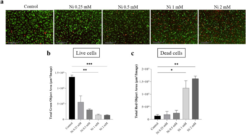
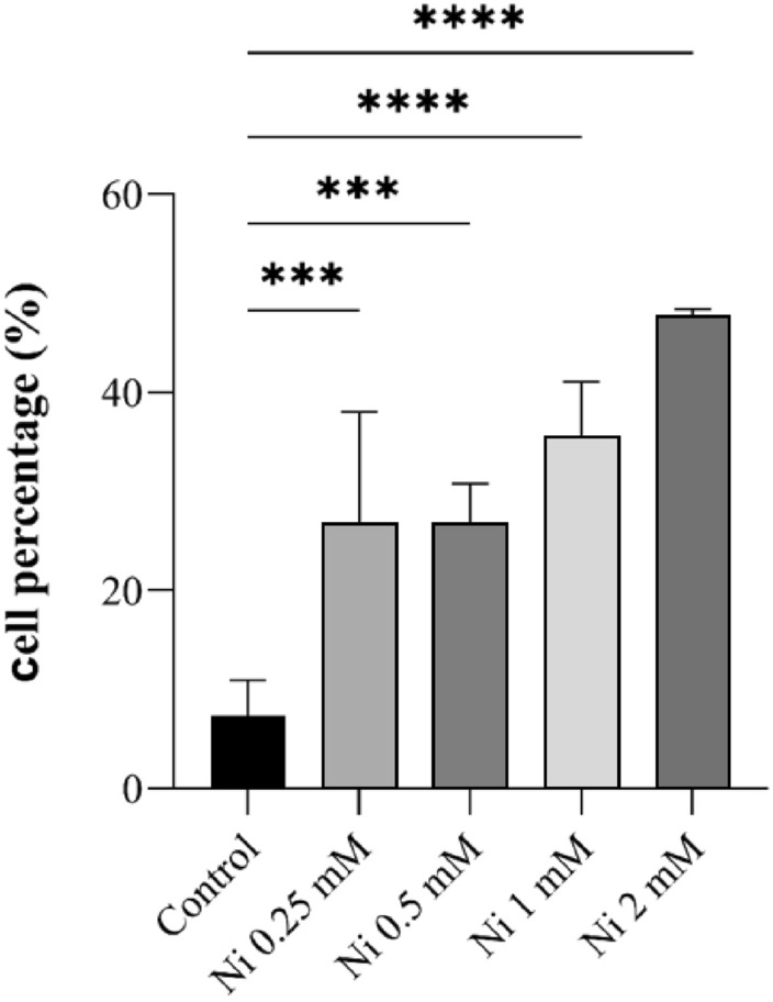
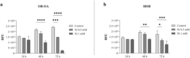
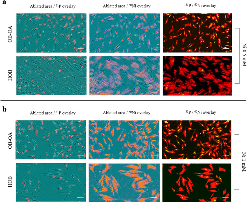
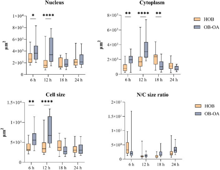
Nickel(Ni)-containing materials have been widely used in a wide range of medical applications, including orthopaedics. Despite their excellent properties, there is still a problem with the release of nickel ions into the patient's body, which can cause changes in the behaviour of surrounding cells and tissues. This study aims to evaluate the effects of Ni on bone cells with an emphasis on the determination of Ni localization in cellular compartments in time. For these purposes, one of the most suitable models for studying the effects induced by metal implants was used-the patient's osteoarthritic cells. Thanks to this it was possible to simulate the pathophysiological conditions in the patient's body, as well as to evaluate the response of the cells which come into direct contact with the material after the implantation of the joint replacement. The largest differences in cell viability, proliferation and cell cycle changes occurred between Ni 0.5 mM and 1 mM concentrations. Time-dependent localization of Ni in cells showed that there is a continuous transport of Ni ions between the nucleus and the cytoplasm, as well as between the cell and the environment. Moreover, osteoarthritic osteoblasts showed faster changes in concentration and ability to accumulate more Ni, especially in the nucleus, than physiological osteoblasts. The differences in Ni accumulation process explains the higher sensitivity of patient osteoblasts to Ni and may be crucial in further studies of implant-derived cytotoxic effects.

Keywords: Implant debris; Laser ablation; Metal distribution; Metal uptake; Nickel; Osteoblasts.


Conflict of interest statement

The authors declare no competing interests.

Figures

Figure 1
Figure 1
Analysis of Ni cytotoxicity after 24 h of cultivation with osteoblasts: (a) representative fluorescent images of green stained live cells and red-stained dead cells in presence of Ni (0.25 mM–2 mM) and in control sample (without Ni). Total green (b) and red (c) object area (µm2/Image) was calculated to determine cytotoxic effects on cells (*p < 0.05, **p < 0.01, ***p < 0.001).
Figure 2
Figure 2
Percentage of dead cells in samples without (control) and with 0.25 mM, 0.5 mM, 1 mM and 2 mM of Ni concentrations. Statistical analysis was performed to reveal the differences in %cells between different Ni concentrations (***p < 0.001, ****p < 0.0001).
Figure 3
Figure 3
Measurement of cell proliferation with/without (control) Ni: (a) measurement of metabolic activity of OB-OA cultured with 0.5 mM and 1 mM of Ni for 24 h, 48 h and 72 h, (b) measurement of metabolic activity of HOB cultured with 0.5 mM and 1 mM of Ni for 24 h, 48 h and 72 h. Statistical analysis was performed with *p < 0.05, **p < 0.01, ***p < 0.001, ****p < 0.0001.
Figure 4
Figure 4
Distribution of 31P and 60Ni in HOB and OB-OA cells at Ni2+ concentrations of 0.5 mM (a) and 1 mM (b). Overlays of ablated 31P and 60Ni with IncuCyte bright field image of cells are shown as ablated area/31P overlay and ablated area/60Ni overlay, respectively. On maps of 31P/60Ni overlay, the presence of 31P is shown in green, 60Ni in red, 31P + 60Ni overlapping in yellow. Scale bar = 100 µm.
Figure 5
Figure 5
Distribution of Ni in HOB and OB-OA loaded with 0.5 mM and 1 mM concentrations measured after 24 h of cultivation with Ni2+ . Results are expressed as nucleus/cytoplasm ratio calculated from 60Ni intensities.
Figure 6
Figure 6
31P (green) and 60Ni (red) distribution in HOB and OB-OA after 6 h, 12 h, 18 h and 24 h of cultivation with Ni. 31P and 60Ni overlapping is presented in yellow. Control sample (cells without Ni) was added to prove the absence of Ni.
Figure 7
Figure 7
Analysis of HOB and OB-OA area of the cell, nucleus and cytoplasm and nucleus/cytoplasm (N/C) size ratio calculated from the ablation maps. Statistical analysis of the difference between two cell lines were performed in the time points of 6 h, 12 h, 18 h and 24 h.
Figure 8
Figure 8
Ni distribution in cell expressed as nucleus/cytoplasm ratio in HOB, OB-OA cells, and the comparison of Ni distribution between HOB and OB-OA cell lines: (a) total cell intensity, (b) mean intensity/pixel. Statistical analysis was performed to examine if there are differences between different time points and cell lines (*p < 0.05, **p < 0.01, ***p < 0.001, ****p < 0.0001).
Figure 9
Figure 9
Schematic representation of 60Ni summary intensities (in cps) in the cell compartments (nucleus and cytoplasm) for HOB and OB-OA in time (6 h, 12 h, 18 h and 24 h). Intensity of a blue colouring also reflects Ni concentration in nucleus and cytoplasm.

Similar articles

Cited by

References

    1. Cempel M, Nikel G. Nickel: A review of its sources and environmental toxicology. Polish J. Environ. Stud. 2006;15:375–382.
    1. Henderson RG, Durando J, Oller AR, Merkel DJ, Marone PA, Bates HK. Acute oral toxicity of nickel compounds. Regul. Toxicol. Pharmacol. 2012;62:425–432. doi: 10.1016/j.yrtph.2012.02.002. - DOI - PubMed
    1. Radev DD. Nickel-containing alloys for medical application obtained by methods of mechanochemistry and powder metallurgy. ISRN Metall. 2012;2012:1–6. doi: 10.5402/2012/464089. - DOI
    1. Ahlström MG, Thyssen JP, Wennervaldt M, Menné T, Johansen JD. Nickel allergy and allergic contact dermatitis: A clinical review of immunology, epidemiology, exposure, and treatment. Contact Dermat. 2019;81:227–241. doi: 10.1111/cod.13327. - DOI - PubMed
    1. Keegan GM, Learmonth ID, Case CP. Orthopaedic metals and their potential toxicity in the arthroplasty patient. A review of current knowledge and future strategies. J. Bone Jt. Surg.—Ser Br. 2007;89:567–573. doi: 10.1302/0301-620X.89B5.18903. - DOI - PubMed

Publication types

LinkOut - more resources