. 2024 Jul 12;7(1):855.
doi: 10.1038/s42003-024-06553-3.

A type II phosphatidylinositol-4-kinase coordinates sorting of cargo polarizing by endocytic recycling

Affiliations

A type II phosphatidylinositol-4-kinase coordinates sorting of cargo polarizing by endocytic recycling

Anezia Kourkoulou et al. Commun Biol. .

Abstract

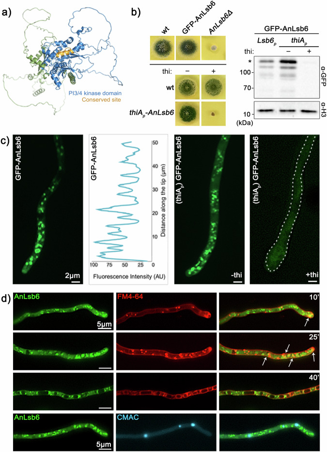
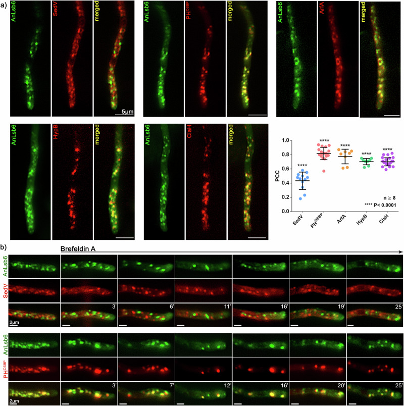
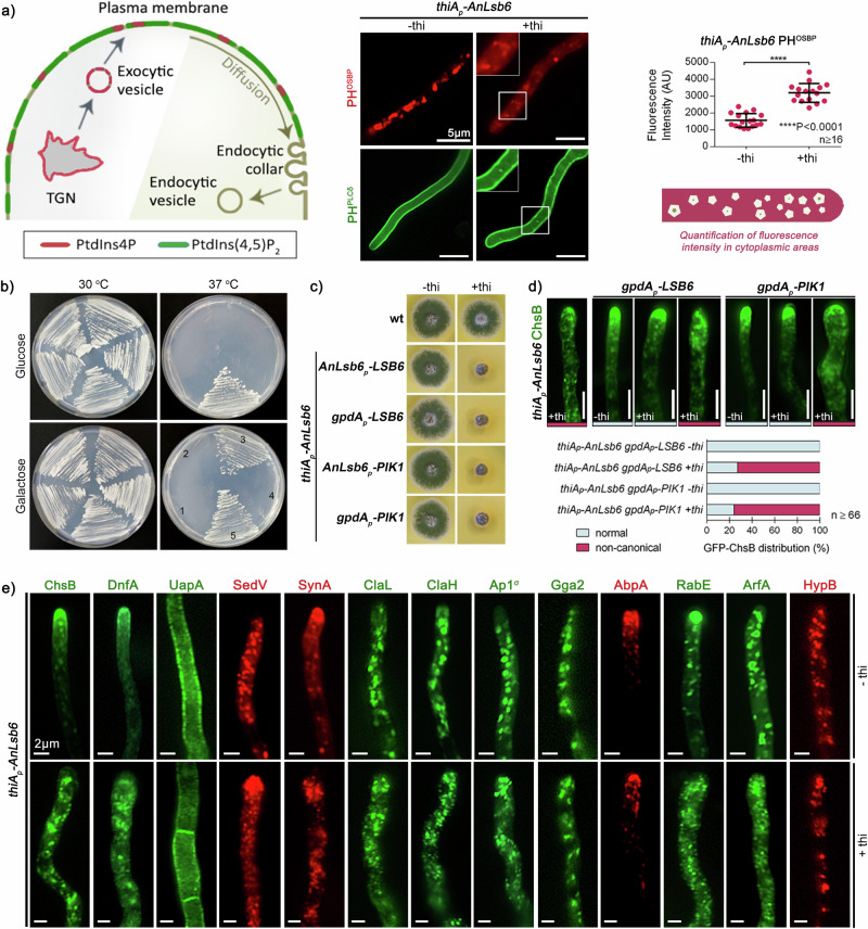
Depending on their phosphorylation status, derivatives of phosphatidylinositol play important roles in vesicle identity, recognition and intracellular trafficking processes. In eukaryotic cells, phosphatidylinositol-4 phosphate pools generated by specific kinases are key determinants of the conventional secretion pathways. Earlier work in yeast has classified phosphatidylinositol-4 kinases in two types, Stt4p and Pik1p belonging to type III and Lsb6p to type II, with distinct cellular localizations and functions. Eurotiomycetes appear to lack Pik1p homologues. In Aspergillus nidulans, unlike homologues in other fungi, AnLsb6 is associated to late Golgi membranes and when heterologously overexpressed, it compensates for the thermosensitive phenotype in a Saccharomyces cerevisiae pik1 mutant, whereas its depletion leads to disorganization of Golgi-associated PHOSBP-labelled membranes, that tend to aggregate dependent on functional Rab5 GTPases. Evidence provided herein, indicates that the single type II phosphatidylinositol-4 kinase AnLsb6 is the main contributor for decorating secretory vesicles with relevant phosphatidylinositol-phosphate species, which navigate essential cargoes following the route of apical polarization via endocytic recycling.


Conflict of interest statement

The authors declare no competing interests.

Figures

Fig. 1
Fig. 1. AnLsb6 is essential for growth and localizes in polarly distributed structures.
a Model of AnLsb6 generated with AlphaFold2. The structure is colored based on domain and conserved site topology, as predicted by InterPro analysis. For coloring based on Predicted Aligned Error (PAE), see Supplementary Fig. 1. The conserved site corresponds to residues 385ELEKLVILDYIMRNTDRGLDN405, part of which (397RNTDRGLDN405) is predicted to operate as a catalytic loop (PROSITE entry PS00916). b Growth of strains carrying deletions of AnLsb6 ORF (ANlsb6Δ), N-terminal GFP-tagged version of AnLsb6 and isogenic strains carrying in locus thi-repressible alleles of thiAp-AnLsb6 in the absence (−) or presence (+) of thiamine (thi). Growth was assessed on minimal media, pH 6.8, 37o C for 72 h. Western blot analysis comparing protein levels of GFP-AnLsb6 and GFP-AnLsb6 expressed under the thiAp promoter in the absence (−) or presence (+) of thiamine (thi). Equal loading is indicated by Histone 3 levels. An asterisk (*) indicates the intact GFP-AnLsb6 protein. Comparison of N- and C-terminal GFP-tagged versions of AnLsb6 and uncropped images of the blots are shown in Supplementary Fig. 2. c Localization of strains expressing N- and C-terminal tagged versions of AnLsb6. A strain repressed for GFP-AnLsb6 expression ( + thi, right panel) is also shown as a control of thiamine repression. Raw data on the quantification of fluorescent intensity are given in Supplementary Data 1. d Localization of GFP-AnLsb6 in the presence of FM4-64, which dynamically labels endocytic steps (plasma membrane, early and late endosomes, vacuoles) and in the presence of the vacuolar stain 7-amino-4-chloromethylcoumarin (Blue CMAC). A minor overlap is observed at earlier time points of the FM4-64 chase (indicated by arrows), in agreement with the non-association with CMAC, that mostly stains terminal endocytic stages.
Fig. 2
Fig. 2. AnLsb6 associates with Golgi structures.
a Localization of GFP-AnLsb6 compared to mRFP- or mCherry-tagged early- and late-Golgi markers. AnLsb6 associates significantly with late-Golgi markers like the pleckstrin homology domain of the human oxysterol binding protein PHOSBP (PCC = 0.84, P < 0.0001, n = 18), GTPase ArfAARF1 (PCC = 0.78, P < 0.0001, n = 8), GEF HypBSEC7 (PCC = 0.72, P < 0.0001, n = 9) and clathrin heavy chain ClaH (PCC = 0.71, P < 0.0001, n = 28), but much less with the early Golgi t-SNARE SedVSED5 (PCC = 0.43, P < 0.0001, n = 12). Raw data on the quantification of co-localization by calculating Pearson’s Correlation Coefficient (PCC) are given in Supplementary Data 2. See Methods for statistical analysis and statistical tests used. Error bars represent Standard Deviation (SD). b Representative images in different hyphae of the subcellular localization of AnLsb6 with SedVSED5 or the pleckstrin homology domain of the human oxysterol binding protein PHOSBP in the presence of the inhibitor Brefeldin A, chased for a time period of 25 min. Notice that a fraction of Brefeldin bodies primarily includes PHOSBP and not SedVSED5.
Fig. 3
Fig. 3. AnLsb6 affects the action of PtdIns4-specific sensors and heterologously complements thermosensitivity in a PIK1Δ mutant.
a Cellular distribution of PtdIns4P and PtdIns(4,5)P2 as monitored by the fluorescence of the PtdIns4P- and PtdIns(4,5)P2-specific sensors, the pleckstrin homology domains of the oxysterol binding protein (PHOSBP) and the rat phospholipase C1 (PHPLCδ), in conditions of AnLsb6 depletion ( + thi). Whereas PHPLCδ localization remains largely undisturbed at the plasma membrane with the exception of a few scattered spots, an almost complete disorganization of the polarized structures recognized by PHOSBP and an increase of cytoplasmic haze (2.04-fold; P < 0.0001; -thi n = 17, +thi n = 16) is observed in the case of PHOSBP. Raw data on the quantification of fluorescent cytoplasmic areas are given in Supplementary Data 3. See Methods for statistical analysis and statistical tests used. Error bars represent SD. b Growth of S. cerevisiae PIK1Δ strains expressing the empty vectors pFL038 (Nr. 1) and pCG563 (Nr. 2), AnLsb6 driven under the yeast PIK1 promoter (Nr. 4) or overexpressed under the galactose responsive galactokinase promoter GAL1 (Nr. 3), compared to the wt (Nr. 5) in the presence of appropriate carbon sources (2% Glucose, 2% Galactose). Growth was assessed on minimal media, at 30o and 37o C for 96 h. c Growth test of LSB6 and PIK1 expressed under the native Anlsb6 promoter or overexpressed under the glyceraldehyde-3-phosphate dehydrogenase promoter gpdA, at conditions where AnLsb6 is expressed (-thi) or repressed by thiamine ( + thi). Growth was assessed on minimal media, pH 6.8, 37o C for 72 h. Fluorescence microscopy and quantification of ChsB rescue for the strains harboring LSB6 and PIK1 under the gpdA promoter is shown in (d). The wt GFP-tagged ChsB resembles the condition where AnLsb6 is expressed in the gpdAp-LSB6 and gpdAp-PIK1 strains (for a -thi GFP-ChsB control see Fig. 3e). Colors: Light gray, normal; Dark pink, non-canonical. For quantification of thiAp-AnLsb6 GFP-ChsB +thi, see also Supplementary Fig. 5. Raw data on the quantification of growth are given in Supplementary Data 5 (thiAp-AnLsb6 gpdAp-LSB6 -thi n = 172; thiAp-AnLsb6 gpdAp-LSB6 +thi n = 108; thiAp-AnLsb6 gpdAp-PIK1 -thi n = 66; thiAp-AnLsb6 gpdAp-PIK1 +thi n = 112). e Cellular localization of GFP- or mRFP/mCherry-tagged protein cargos under conditions where AnLsb6 is expressed (upper panel) or repressed by thi (lower panel, +thi). The cargoes tested are the chitin synthase ChsB, the flippase DnfA, the transporter UapA, the t-SNARE SedVSED5, the synaptobrevin homologue SynA, clathrin light and heavy chains ClaL and ClaH, the adaptors Ap1σ and Gga2, the actin related protein AbpAABP1, the Rab GTPase RabERAB11, the GTPase ArfAARF1 and the GEF HypBSEC7. Notice the misplacement or disorganization of most proteins, with the exception of the plasma membrane transporter UapA that is largely unaffected.
Fig. 4
Fig. 4. AnLsb6 repression leads to RabA/BRAB5-dependent endosomal arrest of the chitin synthase ChsB.
a Speculative scheme of components of the endocytic recycling pathway at the hyphal tip, adopted and modified from Hernández-González et al.. Subcellular localization of ChsB in strains carrying single thiA-rabC, thiA-rabE, and thiA-rabA at repressive conditions in the presence of thiamine ( + thi), as well as in a genetic background where rabB is deleted. b Localization of ChsB in the presence of FM4-64 and of the vacuolar stain CMAC. c Colocalization and relative quantification of GFP-ChsB with mRFP-tagged versions of RabA and RabB under conditions where AnLsb6 is expressed (-thi) or repressed by thiamine ( + thi) (RabA-ChsB: -thi PCC = 0.33 n = 12, +thi PCC = 0.80 n = 13, P < 0.0001; RabB-ChsB: -thi PCC = 0.67 n = 12, +thi PCC = 0.83 n = 10, P = 0.0093; RabA-CMAC PCCA = 0.48 n = 12, RabB-CMAC PCCB = 0.68 n = 10, ChsB-CMAC PCCC = 0.54 n = 22, PCCA-PCCB P = 0.0002, PCCB-PCCC P = 0.0050). Raw data on the quantification of co-localization by calculating Pearson’s Correlation Coefficient (PCC) are given in Supplementary Data 8. Error bars represent SD. See Methods for statistical analysis and statistical tests used. d Growth of single and double mutants of thiA-rabA, rabBΔ and subcellular localization of apical cargos (ChsB, SynA) in rabBΔ or thiA-rabA genetic backgrounds and at conditions where AnLsb6 is expressed (-thi) or repressed by thiamine ( + thi). Localization of ChsB in a rabBΔ background and at conditions where both AnLsb6, RabA and RabC are repressed by thiamine ( + thi). e Subcellular localization of ChsB in a strain carrying a single thiA-slaB allele in the absence or presence of thiamine, and at conditions where both AnLsb6 and SlaBSLA2 are either expressed (-thi) or repressed by thiamine ( + thi).

Similar articles

Cited by

References

    1. Strahl T, Thorner J. Synthesis and function of membrane phosphoinositides in budding yeast, Saccharomyces cerevisiae. Biochim. Biophys. Acta. 2007;1771:353–404. doi: 10.1016/j.bbalip.2007.01.015. - DOI - PMC - PubMed
    1. Posor Y, Jang W, Haucke V. Phosphoinositides as membrane organizers. Nat. Rev. Mol. Cell. Biol. 2022;23:797–816. doi: 10.1038/s41580-022-00490-x. - DOI - PMC - PubMed
    1. Balla T. Phosphoinositides: tiny lipids with giant impact on cell regulation. Physiol. Rev. 2013;93:1019–1137. doi: 10.1152/physrev.00028.2012. - DOI - PMC - PubMed
    1. Di Paolo G, De Camilli P. Phosphoinositides in cell regulation and membrane dynamics. Nature. 2006;443:651–657. doi: 10.1038/nature05185. - DOI - PubMed
    1. Jean S, Kiger AA. Coordination between RAB GTPase and phosphoinositide regulation and functions. Nat. Rev. Mol. Cell. Biol. 2012;13:463–470. doi: 10.1038/nrm3379. - DOI - PubMed

MeSH terms

Substances

LinkOut - more resources