. 2024 Jul 13;24(1):784.
doi: 10.1186/s12903-024-04518-w.

Quality of different obturation techniques to fill perforating internal root resorption: a micro-computed tomographic study

Affiliations

Quality of different obturation techniques to fill perforating internal root resorption: a micro-computed tomographic study

Shaimaa Mohamed Abu El Sadat et al. BMC Oral Health. .

Abstract

Background: This study aimed to assess the quality of various obturation techniques to fill perforation caused by internal root resorption using Micro-computed Tomography.

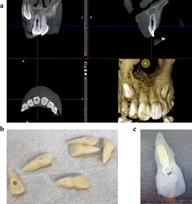
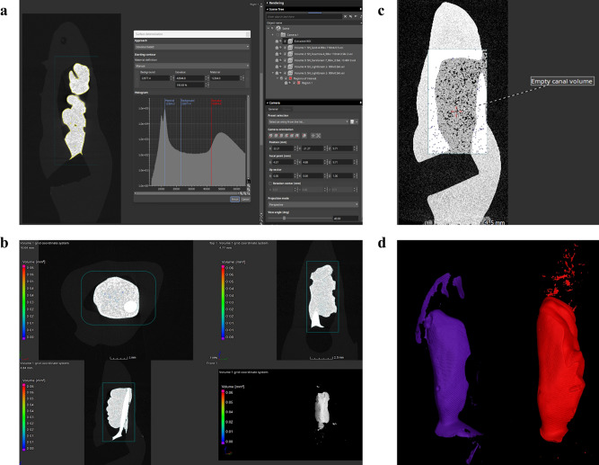
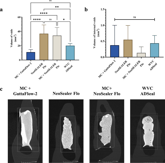
Methods: Cone-beam computed tomography images of a maxillary central incisor tooth with perforating internal resorptive defect were used to create a 3D printed model of the affected tooth. The replicas were divided into four groups based on the obturation technique used. The techniques included Group 1: a polydimethylsiloxane-based sealer (GuttaFlow-2) with gutta-percha. Group 2: same as Group 1 except for using a pre-mixed Bioceramic-based sealer (NeoSEALER Flo). Group 3: the defect was filled entirely using the NeoSealer Flo Bioceramic-based sealer. Group 4: the samples were obturated using the warm vertical compaction technique with a resin-based sealer (ADSeal). The resin models were then scanned a micro-computed scanner to evaluate the percentage of voids in each group.

Results: The results showed that NeoSEALER Flo groups had significantly the highest volume of voids while GuttaFlow-2 and warm vertical compaction groups had the lowest void volume.

Conclusions: GuttaFlow-2 and warm vertical compaction techniques performed best in filling the internal resorptive defect.

Keywords: GuttaFlow-2; Internal Root Resorption; Micro-CT; NeoSealer Flo; Root canal Obturation; Root canal perforation.


Conflict of interest statement

The authors declare no competing interests.

Figures

Fig. 1
Fig. 1
(a). High-resolution CBCT scan showing the internal resorption defect. (b). 3D printed teeth with virtual access cavity. (c). Calcium hydroxide paste injected and radiographed to check the radiographic appearance of the resin material
Fig. 2
Fig. 2
(a) Surface determination of the filled material with the histogram of the ROI. (b). MPR screen displaying the internal porosities of the bioceramic sealer with a single cone technique. (c). Volume calculation of empty canal. (d). 3D volume representation of the filling material, the purple volume is for the gutta flow and gutta percha while the red volume is for the bioceramic sealer only
Fig. 3
Fig. 3
(a). Bar graphs showing the volume of voids between the different groups. (b). Bar graphs showing the volume of internal defects between the different groups. (c). Microcomputed tomography representative images showing the canal filling of the different groups. Data are presented as mean ± standard deviations. MC; master cone, WVC; warm vertical condensation, ns; non-significant, *p < 0.05, **p < 0.01, ****p < 0.0001

Similar articles

Cited by

References

    1. Fernandes M, de Ataide I, Wagle R. Tooth resorption part I - pathogenesis and case series of internal resorption. J Conserv Dent. 2013;16:4–8. doi: 10.4103/0972-0707.105290. - DOI - PMC - PubMed
    1. Patel S, Ricucci D, Durak C, Tay F. Internal root resorption: a review. J Endod. 2010;36:1107–21. doi: 10.1016/j.joen.2010.03.014. - DOI - PubMed
    1. Rossi-Fedele G, Figueiredo JA, Abbott PV. Teeth with double internal inflammatory resorption: report of two cases. Aust Endod J. 2010;36:122–9. doi: 10.1111/j.1747-4477.2009.00197.x. - DOI - PubMed
    1. Collins J, Walker MP, Kulild J, Lee C. A comparison of three gutta-percha obturation techniques to replicate canal irregularities. J Endod. 2006;32:762–5. doi: 10.1016/j.joen.2005.10.001. - DOI - PubMed
    1. Kulild J, Lee C, Dryden J, Collins J, Feil P. A comparison of 5 gutta-percha obturation techniques to replicate canal defects. Oral Surg Oral Med Oral Pathol Oral Radiol Endod. 2007;103:e28–32. doi: 10.1016/j.tripleo.2006.06.007. - DOI - PubMed

MeSH terms

LinkOut - more resources