. 2024 Jul 13;24(1):785.
doi: 10.1186/s12903-024-04549-3.

Effect of autophagy on aging-related changes in orthodontic tooth movement in rats

Affiliations

Effect of autophagy on aging-related changes in orthodontic tooth movement in rats

Bowen Xu et al. BMC Oral Health. .

Abstract

Background: The number of adult orthodontic patients is increasing, and studies have shown that autophagy is involved in regulating orthodontic tooth movement and plays an important role in aging-related changes. Therefore, we aimed to explore the role of autophagy in aging-related changes during orthodontic tooth movement by establishing a rat orthodontic tooth movement model.

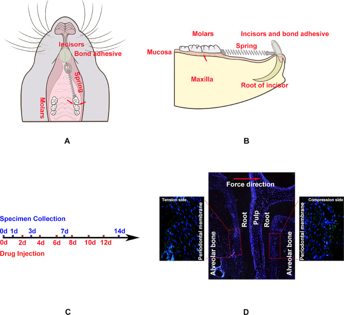
Methods: Forty-five 6-week-old and sixty-five 8-month-old male Sprague-Dawley rats were selected to represent adolescents and adults and establish orthodontic tooth movement model. They were sacrificed on days 0,1,3,7 and 14. Immunohistochemistry, immunofluorescence and tartrate resistant acid phosphatase (TRAP) staining were applied to measure the expression level of osteogenesis, autophagy, aging factors and osteoclast number in periodontal membrane of left upper first molar during orthodontic tooth movement. Then, we regulated the autophagy level by injecting autophagy activator rapamycin during orthodontic tooth movement and measured these factors and tooth movement distance by micro-computed tomography.

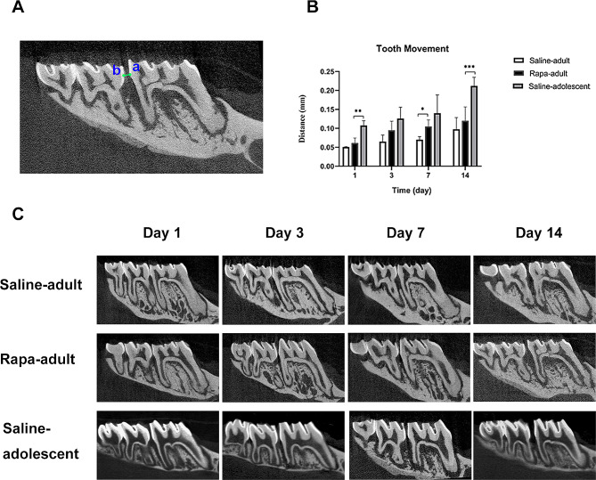
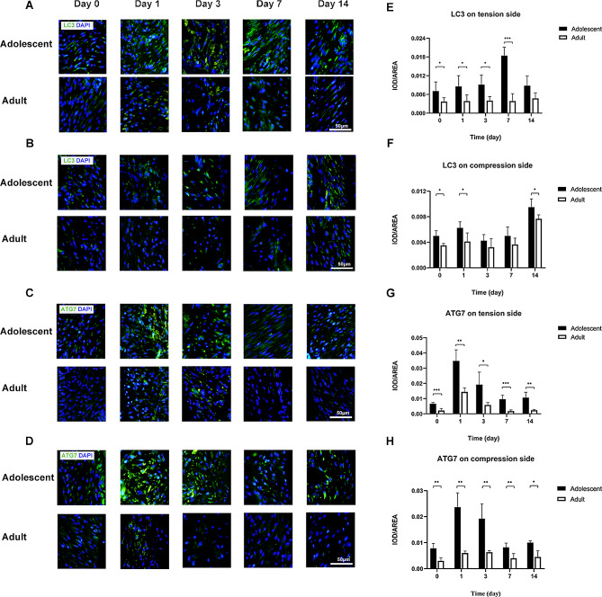
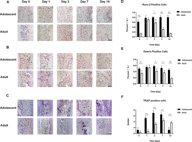
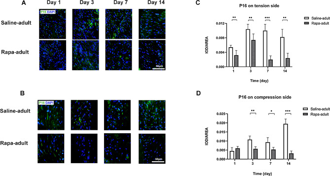
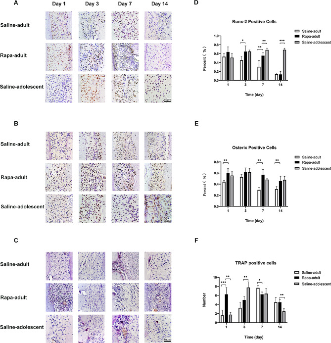
Results: Aging factor levels in the periodontal membrane were higher in adult rats than in adolescent rats and the autophagy factor levels were lower. The levels of osteogenic factors were lower on the tension side in adult rats than in adolescent rats. The peak osteoclast number on the pressure side occurred later in adult rats than in adolescent rats. The injection of rapamycin increased autophagy, accelerated orthodontic tooth movement in adult rats, and reduced the levels of aging factors. The levels of osteogenic factors were higher and reached those in adolescent rats at some time points. The number of osteoclasts increased significantly in the early stage.

Conclusions: Autophagy may play a substantial role in regulating aging-related changes in orthodontic tooth movement.

Keywords: Aging; Autophagy; Bone remodeling; Tooth movement.


Conflict of interest statement

The authors declare no competing interests.

Figures

Fig. 1
Fig. 1
Illustration of the experimental methods A, B Diagram of orthodontic tooth movement model establishment and drug injection. Arrows show the drug injection points C Flowchart of drug injection and specimen collection D Diagram of the zone of interest for histopathological analysis on the tension side and compression side of the periodontal ligament of the left maxillary first molar
Fig. 2
Fig. 2
Orthodontic tooth movement in saline-adult, rapa-adult and saline-adolescent rats A Method of orthodontic tooth movement evaluation. Point a refers to the most distal point of the crown of the maxillary left first molar, and point b refers to the most mesial point of the crown of the second molar. The distance of line ab represents the orthodontic tooth movement distance B Comparison of the orthodontic tooth movement distance between the saline-adult, rapa-adult and saline-adolescent rats. (n = 5, *P < 0.05, **P < 0.01, ***P < 0.001) C Comparison of orthodontic tooth movement between the saline-adult, rapa-adult and saline-adolescent rats on micro-computed tomography
Fig. 3
Fig. 3
Autophagy level on tension and compression side of periodontal membrane in adolescent and adult rats A LC3 in the periodontal membrane on the tension side in adolescent and adult rats by immunofluorescence staining, with LC3 in green and nuclei in blue B LC3 in the periodontal membrane on the compression side in adolescent and adult rats by immunofluorescence staining, with LC3 in green and nuclei in blue. C ATG7 in the periodontal membrane on the tension side in adolescent and adult rats by immunofluorescence staining, with ATG7 in green and nuclei in blue D ATG7 in the periodontal membrane on the compression side in adolescent and adult rats by immunofluorescence staining, with ATG7 in green and nuclei in blue. Scale bar indicates 50 μm E Expression level of LC3 in the periodontal membrane on the tension side in adolescent and adult rats F Expression level of LC3 in the periodontal membrane on the com-pression side in adolescent and adult rats G Expression level of ATG7 in the periodontal membrane on the tension side in adolescent and adult rats H Expression level of ATG7 in the periodontal membrane on the compression side in adolescent and adult rats. (n = 5, *P < 0.05, **P < 0.01, ***P < 0.001)
Fig. 4
Fig. 4
P16 level on tension and compression side of periodontal membrane in adolescent and adult rats A P16 in the periodontal membrane on the tension side in adolescent and adult rats by immunofluorescence staining, with P16 in green and nuclei in blue B P16 in the periodontal membrane on the compression side between adolescent and adult rats by immunofluorescence staining, with P16 in green and nuclei in blue. Scale bar indicates 50 μm C Expression level of P16 in the periodontal membrane on the tension side in adolescent and adult rats D Expression level of P16 in the periodontal membrane on the compression side in adolescent and adult rats. (n = 5, *P < 0.05, **P < 0.01, ***P < 0.001)
Fig. 5
Fig. 5
Osteogenesis level and osteoclast number in periodontal membrane in adolescent and adult rats A Runx-2-positive cells in the periodontal membrane on the tension side in adolescent and adult rats by immunohistochemistry B Osterix-positive cells in the periodontal membrane on the tension side in adolescent and adult rats by immunohistochemistry C Mature osteoclasts stained by TRAP (red, with multiple nuclei) in the periodontal membrane on the compression side in adolescent and adult rats. Scale bar indicates 50 μm D Percentage of Runx-2-positive cells in the periodontal membrane on the tension side in adolescent and adult rats E Percentage of Osterix-positive cells in the periodontal membrane on the tension side in adolescent and adult rats F Number of mature osteoclasts in the periodontal membrane on the compression side in adolescent and adult rats. (n = 5, *P < 0.05, **P < 0.01, ***P < 0.001)
Fig. 6
Fig. 6
Autophagy level on tension and compression side of periodontal membrane in saline-adult and rapa-adult rats A LC3 in the periodontal membrane on the tension side in saline-adult and rapa-adult rats by immunofluorescence staining, with LC3 in green and nuclei in blue B LC3 in the periodontal membrane on the compression side in saline-adult and rapa-adult rats by immunofluorescence staining, with LC3 in green and nuclei in blue C ATG7 in the periodontal membrane on the tension side in saline-adult and rapa-adult rats by immunofluorescence staining, with in ATG7 green and nuclei in blue D ATG7 in the periodontal membrane on the compression side in saline-adult and rapa-adult rats by immunofluorescence staining, with ATG7 in green and nuclei in blue. Scale bar indicates 50 μm E Expression level of LC3 in the periodontal membrane on the tension side in saline-adult and rapa-adult rats F Expression level of LC3 in the periodontal membrane on the compression side in saline-adult and rapa-adult rats G Expression level of ATG7 in the periodontal membrane on the tension side in saline-adult and rapa-adult rats H Expression level of ATG7 in the periodontal membrane on the compression side in saline-adult and rapa-adult rats. (n = 5, *P < 0.05, **P < 0.01, ***P < 0.001)
Fig. 7
Fig. 7
P16 level on tension and compression side of periodontal membrane in saline-adult and rapa-adult rats A P16 in the periodontal membrane on the tension side in saline-adult and rapa-adult rats by immunofluorescence staining, with P16 in green and nuclei in blue. B P16 in the periodontal membrane on the compression side in saline-adult and rapa-adult rats by immunofluorescence staining, with P16 in green and nuclei in blue. Scale bar indicates 50 μm C Expression level of P16 in the periodontal membrane on the tension side in saline-adult and rapa-adult rats D Expression level of P16 in the periodontal membrane on the compression side in saline-adult and rapa-adult rats. (n = 5, *P < 0.05, **P < 0.01, ***P < 0.001)
Fig. 8
Fig. 8
Osteogenesis level and osteoclast number in periodontal membrane in saline-adult, rapa-adult and saline-adolescent rats A Runx-2-positive cells in the periodontal membrane on the tension side in saline-adult, rapa-adult and saline-adolescent rats by immunohistochemistry B Osterix-positive cells in the periodontal membrane on the tension side in saline-adult, rapa-adult and saline-adolescent rats by immunohistochemistry C Mature osteoclasts stained by TRAP (red, with multiple nuclei) in the periodontal membrane on the compression side in saline-adult, rapa-adult and saline-adolescent rats. Scale bar indicates 50 μm D Percentage of Runx-2 positive cells in the periodontal membrane on the tension side in saline-adult, rapa-adult and saline-adolescent rats E Percentage of Osterix-positive cells in the periodontal membrane on the tension side in saline-adult, rapa-adult and saline-adolescent rats F Number of mature osteoclasts in the periodontal membrane on the compression side in saline-adult, rapa-adult and saline-adolescent rats. (n = 5, *P < 0.05, **P < 0.01, ***P < 0.001)

Similar articles

Cited by

References

    1. Zheng J, Xu B, Yang K. Autophagy regulates osteogenic differentiation of Human Periodontal Ligament Stem cells Induced by Orthodontic Tension. Stem Cells Int. 2022;2022:2983862. doi: 10.1155/2022/2983862. - DOI - PMC - PubMed
    1. Zhang Y, Cui Y, Wang L, Han J. Autophagy promotes osteoclast podosome disassembly and cell motility athrough the interaction of kindlin3 with LC3. Cell Signal. 2020;67:109505. doi: 10.1016/j.cellsig.2019.109505. - DOI - PubMed
    1. Kou Y, Li C, Yang P, The W9 peptide inhibits osteoclastogenesis and osteoclast activity by downregulating osteoclast autophagy and promoting osteoclast apoptosis. J Mol Histol., Luther L et al. F. Adults seeking orthodontic treatment: expectations, periodontal and TMD issues. Br Dent J. 2015;218(3):111–117. - PubMed
    1. L., Christensen F., Luther (2015) Adults seeking orthodontic treatment: expectations periodontal and TMD issues British Dental Journal 218(3) 111-117 10.1038/sj.bdj.2015.46 - PubMed
    1. Xu B, Yang K. Changes in alveolar bone structure during orthodontic tooth movement in adolescent and adult rats: a microcomputed tomography study. Orthod Craniofac Res. 2023;26(4):568–75. doi: 10.1111/ocr.12646. - DOI - PubMed

Substances

LinkOut - more resources