. 2024 Jul 12;21(1):170.
doi: 10.1186/s12974-024-03159-8.

Deletion of myeloid HDAC3 promotes efferocytosis to ameliorate retinal ischemic injury

Affiliations

Deletion of myeloid HDAC3 promotes efferocytosis to ameliorate retinal ischemic injury

Rami A Shahror et al. J Neuroinflammation. .

Abstract

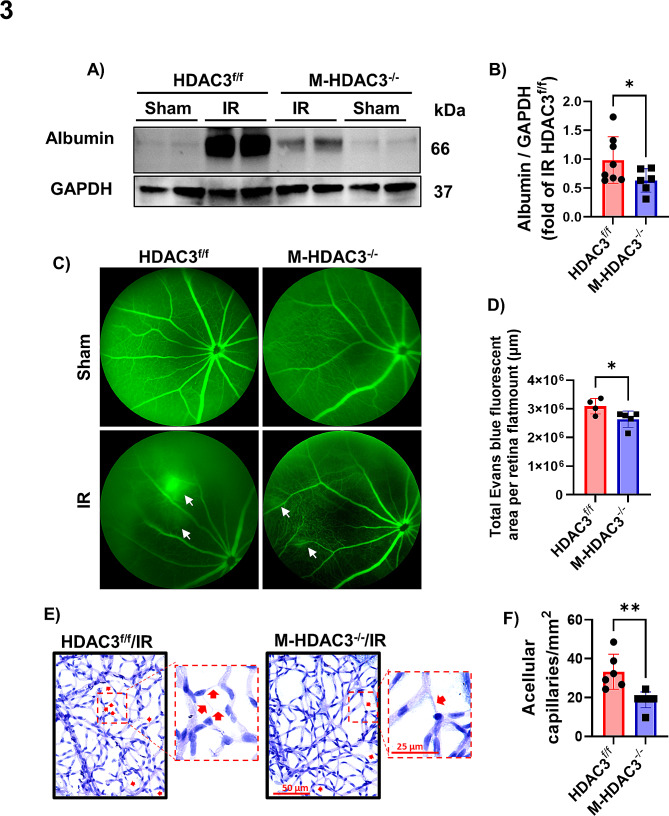
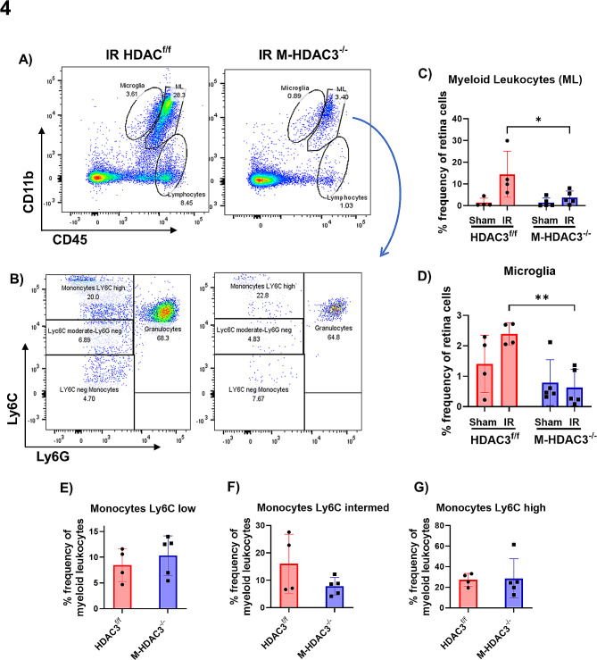
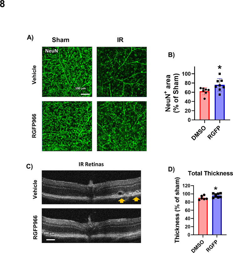
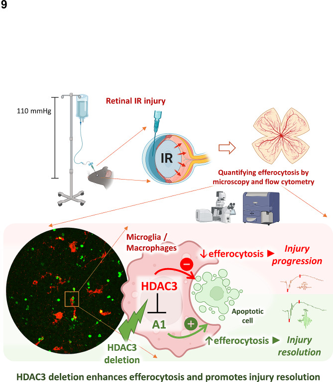
Ischemia-induced retinopathy is a hallmark finding of common visual disorders including diabetic retinopathy (DR) and central retinal artery and vein occlusions. Treatments for ischemic retinopathies fail to improve clinical outcomes and the design of new therapies will depend on understanding the underlying disease mechanisms. Histone deacetylases (HDACs) are an enzyme class that removes acetyl groups from histone and non-histone proteins, thereby regulating gene expression and protein function. HDACs have been implicated in retinal neurovascular injury in preclinical studies in which nonspecific HDAC inhibitors mitigated retinal injury. Histone deacetylase 3 (HDAC3) is a class I histone deacetylase isoform that plays a central role in the macrophage inflammatory response. We recently reported that myeloid cells upregulate HDAC3 in a mouse model of retinal ischemia-reperfusion (IR) injury. However, whether this cellular event is an essential contributor to retinal IR injury is unknown. In this study, we explored the role of myeloid HDAC3 in ischemia-induced retinal neurovascular injury by subjecting myeloid-specific HDAC3 knockout (M-HDAC3 KO) and floxed control mice to retinal IR. The M-HDAC3 KO mice were protected from retinal IR injury as shown by the preservation of inner retinal neurons, vascular integrity, and retinal thickness. Electroretinography confirmed that this neurovascular protection translated to improved retinal function. The retinas of M-HDAC3 KO mice also showed less proliferation and infiltration of myeloid cells after injury. Interestingly, myeloid cells lacking HDAC3 more avidly engulfed apoptotic cells in vitro and after retinal IR injury in vivo compared to wild-type myeloid cells, suggesting that HDAC3 hinders the reparative phagocytosis of dead cells, a process known as efferocytosis. Further mechanistic studies indicated that although HDAC3 KO macrophages upregulate the reparative enzyme arginase 1 (A1) that enhances efferocytosis, the inhibitory effect of HDAC3 on efferocytosis is not solely dependent on A1. Finally, treatment of wild-type mice with the HDAC3 inhibitor RGFP966 ameliorated the retinal neurodegeneration and thinning caused by IR injury. Collectively, our data show that HDAC3 deletion enhances macrophage-mediated efferocytosis and protects against retinal IR injury, suggesting that inhibiting myeloid HDAC3 holds promise as a novel therapeutic strategy for preserving retinal integrity after ischemic insult.

Keywords: Efferocytosis; Histone deacetylase 3; Macrophages; Microglia; Retinal ischemia-reperfusion injury.


Conflict of interest statement

The authors declare no competing interests.

Figures

Fig. 1
Fig. 1
Myeloid HDAC3 deletion is neuroprotective and mitigates retinal thinning after IR injury. (A) Immunolabeling of postmortem human retina sections from control and DR patients show colocalization of HDAC3 with Iba1+ myeloid cells. GCL: ganglion cell layer, INL: inner nuclear layer, ONL: outer nuclear layer, n = 3. (B) Graph extracted from a searchable transcriptome database for human eye tissue (https://www.eye-transcriptome.com/) shows high HDAC3 expression in retinal microglia and vitreous MΦ. C-E) Immunolabeling at 7 days after injury and quantification show reduced neurodegeneration (marked by the neuronal marker, NeuN) and microglia/MΦ numbers (marked by Iba1) in the M-HDAC3−/− retinas as compared to HDAC3f/f, n = 5–9. F-H) Optical coherence tomography (OCT) performed on anesthetized mice 7 days after IR shows preserved total retinal thickness (indicated by a dashed line) and preserved thickness of the inner nuclear layer (indicated by a star) in M-HDAC3−/− retinas, n = 6–9, *p < 0.05, **p < 0.01, ***p < 0.005, ns: not significant
Fig. 2
Fig. 2
Myeloid HDAC3 deletion improves the retinal ERG after IR. A-C) Representative a- and b-waveforms elicited by 3 flash intensities in retinas of HDAC3f/f and M-HDAC3−/− mice on day 14 after IR injury. D-I) Quantification and comparison reveal improved waveform amplitudes in M-HDAC3−/− retinas with statistical significance achieved at flash intensities of 0.1 cd.s/m2 (a-wave) and 0.01 cd.s/m2 and 1.0 cd.s/m² (b-wave). J-L) Representative waveforms of oscillatory potentials (OPs) in retinas of HDAC3f/f and M-HDAC3−/− mice elicited by 3 flash intensities at day 14 after IR injury. M-U) Quantification reveals a pattern of improved OP with statistical significance achieved at 1 cd.s/m² (OP2) and at 0.1 cd.s/m2 and 1.0 cd.s/m² (OP3), n = 6–8, *p < 0.05, **p < 0.01
Fig. 3
Fig. 3
Myeloid HDAC3 deletion is vascular protective in retinal IR injury. A, B) Albumin extravasation at day 2 post-IR was measured in protein lysates of perfused retinas by western blot. Extravasated albumin was markedly reduced in M-HDAC3−/− retinas compared to HDAC3f/f controls and was nearly absent in the contralateral retinas. GAPDH was used as a loading control, n = 6–8. C) Representative retinal fluorescein angiography images at day two post-IR showing increased retinal vascular permeability. Leaked fluorescein demonstrated by diffusely hazy fluorescence (white arrows) was markedly reduced in M-HDAC3−/− retinas compared to HDAC3f/f controls and was absent in the sham retinas. D) Quantification of Evans blue leakage demonstrates a significant reduction in the M-HDAC3−/− IR retinas, n = 4–5. Representative images are included in supplementary figure S8. E, F) Vascular digests at 14 days after IR injury showed increased numbers of acellular retinal capillaries (empty basement membrane sleeves - red arrows) in injured HDAC3f/f retinas compared to injured M-HDAC3−/− retinas, n = 6–8, *p < 0.05, **p < 0.01
Fig. 4
Fig. 4
M-HDAC3 deletion mitigates the IR-induced microglia proliferation and myeloid leukocyte infiltration into the retina. (A) Representative scatter graphs from flow-cytometric analysis showing the initial gating strategy used to quantify the immune cell populations of microglia, myeloid leukocytes (ML), and lymphocytes in HDAC3f/f and M-HDAC3−/− retinas on day two after IR injury. (B) The ML were further gated based on Ly6C/Ly6G expression into Ly6Chi/Ly6Gneg monocytes, Ly6Cmoderate/Ly6Gneg monocytes, Ly6Cneg/Ly6Gneg monocytes, and Ly6C+/Ly6G+ granulocytes. (C) The number of CD11b+/CD45hi ML was reduced in injured M-HDAC3−/− retinas compared to injured HDAC3f/f controls indicating reduced myeloid cell infiltration/proliferation. (D) Similarly, the number of CD11b+/CD45low microglia was reduced in injured M-HDAC3−/− retinas. E-G) ML further gated into Ly6Clow, Ly6Cintermediate, and Ly6Chigh monocytes showed no change in percentage frequency between the injured groups. For each data point, 3 retinas were pooled and n = 4–5 preparations of pooled retinas were analyzed for each group. *p < 0.05, **p < 0.01
Fig. 5
Fig. 5
M-HDAC3 deletion improves macrophage clearance of apoptotic cells through efferocytosisin vivo and in vitro.A) Representative confocal images of immunolabeled TUNEL+ apoptotic cells (red) and Iba1+ myeloid cells (green) in retinal flat mounts on day 2 after IR injury. Arrows and arrowheads depict Iba1-associated and free TUNEL+ cells, respectively. B) The total number of apoptotic cells (ACs, TUNEL+) was not significantly different between the injured groups. C) Quantification of the efferocytosis index (% Iba1+ ACs / total ACs) indicated enhanced phagocytosis in injured retinas of M-HDAC3−/− compared to HDAC3f/f mice, n = 6–8. D-G) Flow-cytometric analysis of myeloid leukocytes and microglia co-expressing the phagocytic marker CD68+ and the apoptotic cell signal PSVue reveals more CD68+/PSVue+ cells in retinas of M-HDAC3−/− compared to HDAC3f/f mice at day 2 after IR injury, n = 5 per group. H) Representative scatter plots from flow cytometry studies in which non-apoptotic (NAC) or apoptotic (AC) CFDA green-labeled K-562 cells were co-cultured with CM-DiI red-labeled MΦ. Control MΦ received no treatment (no ttt). I) Quantification by fluorescence-activated cell sorting (FACS) of MΦ engaged in efferocytosis of ACs (Dil+/CFDA+) as a percent of total MΦ followed by normalization to the NAC group. Efferocytosis was enhanced in HDAC3−/− MΦ, n = 5 per group, *p < 0.05, ***p < 0.005. No-treatment group was used as a negative control for gating of CFDA- macrophages
Fig. 6
Fig. 6
Arginase 1 is upregulated in M-HDAC3−/−macrophages and its inhibition impairs macrophage-mediated efferocytosis. A, B) Western blot shows upregulation of Arginase 1 (A1) in M-HDAC3−/− but not HDAC3f/f MΦ after co-incubation with K-562 ACs. Controls included either co-incubation of MΦ with K-562 NACs or no treatment (no ttt), n = 4. C) The corresponding A1 transcript was increased in M-HDAC3−/− MΦ co-cultured with K-562 ACs as compared to control preparations. A1 transcript also increased in HDAC3f/f MΦ co-cultured with ACs but significantly less than in AC/M-HDAC3−/− MΦ co-cultures, n = 4. D)In vitro efferocytosis assay in which CFDA green-labeled K-562 NACs or ACs (shown in green) were added to DiI red-labeled MΦ (shown in red) after 45 min pre-incubation with the A1 inhibitor ABH (100 µM). Arrowheads indicate efferocytosis of ACs by HDAC3f/f or M-HDAC3−/− MΦ, n = 4. E) Quantification of MΦ engaged in efferocytosis of ACs (Dil+/CFDA+) as a percent of total MΦ reveals enhanced efferocytosis in M-HDAC3−/− MΦ. Two-way ANOVA showed that the effect of HDAC3 deletion and ABH treatment were independently statistically significant but there was no interaction between the two, n = 4, *p < 0.05, **p < 0.01, ***p < 0.005, ****p < 0.001
Fig. 7
Fig. 7
Arginase 1 deletion inhibits efferocytosis in macrophages (A) Representative images from an in vitro efferocytosis assay in which CFDA green-labeled R28 ACs were added to CM-DiI red-labeled MΦ isolated from M-A1−/− or A1f/f mice. (B) Efferocytosis was reduced in MΦ from M-A1−/− mice, n = 3. C, D) A similar efferocytosis assay but using K-562 ACs also showed reduced efferocytosis by MΦ from M-A1−/− mice compared to A1f/f mice, n = 5 per group. E, F) Scatter plots and quantification from FACS of CM-DiI+/CFDA+ MΦ confirmed impaired efferocytosis of K-562 AC by M-A1−/− compared to A1f/f MΦ, n = 5 per group, *p < 0.05, **p < 0.01
Fig. 8
Fig. 8
HDAC3 inhibition is neuroprotective and mitigates retinal thinning after IR injury. A-B) Retinal flat mounts from vehicle-treated WT mice show a marked loss of NeuN-labeled retinal neurons on day 7 after IR injury. After administration of the HDAC3 inhibitor RGFP966 (10 mg/kg i.p.) at 1 h after IR and on days 2, 4, and 6, retinal neurons were partially preserved at 7 days. RGFP996 did not reduce NeuN-positive cells in the retinas of the sham animals, n = 7–8. C, D) OCT images and quantification show preserved total retinal thickness on day 7 after IR injury in WT mice treated with RGFP966. Orange arrows indicate retinal detachment in the IR retinas of vehicle treated group that is absent in the RGFP996 treated group, n = 6–8, *p < 0.05
Fig. 9
Fig. 9
Schematic of the study findings. Myeloid HDAC3 suppresses efferocytosis and A1 expression. A1 promotes efferocytosis and plays a role in the enhanced efferocytosis of MΦ lacking HDAC3. HDAC3 may additionally impair efferocytosis by a mechanism unrelated to loss of A1

References

    1. Hartsock MJ, Cho H, Wu L, Chen WJ, Gong J, Duh EJ. A mouse model of retinal ischemia-reperfusion injury through elevation of intraocular pressure. J Vis Exp. 2016. - PMC - PubMed
    1. Renner M, Stute G, Alzureiqi M, Reinhard J, Wiemann S, Schmid H, et al. Optic nerve degeneration after retinal ischemia/reperfusion in a rodent model. Front Cell Neurosci. 2017;11:254. doi: 10.3389/fncel.2017.00254. - DOI - PMC - PubMed
    1. Ezra-Elia R, Alegro da Silva G, Zanoni DS, Laufer-Amorim R, Vitor Couto do Amaral A, Laus JL, et al. Functional and structural evaluation of sildenafil in a rat model of acute retinal ischemia/reperfusion injury. Curr Eye Res. 2017;42:452–61. doi: 10.1080/02713683.2016.1193615. - DOI - PubMed
    1. Osborne NN, Casson RJ, Wood JP, Chidlow G, Graham M, Melena J. Retinal ischemia: mechanisms of damage and potential therapeutic strategies. Prog Retin Eye Res. 2004;23:91–147. doi: 10.1016/j.preteyeres.2003.12.001. - DOI - PubMed
    1. Shosha E, Shahror RA, Morris CA, Xu Z, Lucas R, McGee-Lawrence ME, et al. The arginase 1/ornithine decarboxylase pathway suppresses hdac3 to ameliorate the myeloid cell inflammatory response: implications for retinal ischemic injury. Cell Death Dis. 2023;14:621. doi: 10.1038/s41419-023-06147-7. - DOI - PMC - PubMed

LinkOut - more resources