. 2024 Jul 12;25(1):273.
doi: 10.1186/s12931-024-02899-8.

The integrin receptor beta7 subunit mediates airway remodeling and hyperresponsiveness in allergen exposed mice

Affiliations

The integrin receptor beta7 subunit mediates airway remodeling and hyperresponsiveness in allergen exposed mice

Miri Assayag et al. Respir Res. .

Abstract

Background: Fibroblast differentiation to a myofibroblast phenotype is a feature of airway remodeling in asthma. Lung fibroblasts express the integrin receptor α4β7 and fibronectin induces myofibroblast differentiation via this receptor.

Objectives: To investigate the role of the β7 integrin receptor subunit and α4β7 integrin complex in airway remodeling and airway hyperresponsiveness (AHR) in a murine model of chronic allergen exposure.

Methods: C57BL/6 wild type (WT) and β7 integrin null mice (β7 -/-) were sensitized (days 1,10) and challenged with ovalbumin (OVA) three times a week for one or 4 weeks. Similar experiments were performed with WT mice in the presence or absence of α4β7 blocking antibodies. Bronchoalveolar (BAL) cell counts, AHR, histological evaluation, soluble collagen content, Transforming growth factor-β (TGFβ) and Interleukin-13 (IL13) were measured. Phenotype of fibroblasts cultured from WT and β7 -/- saline (SAL) and OVA treated mice was evaluated.

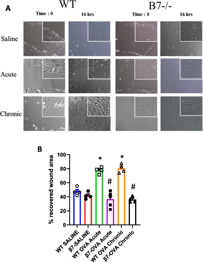
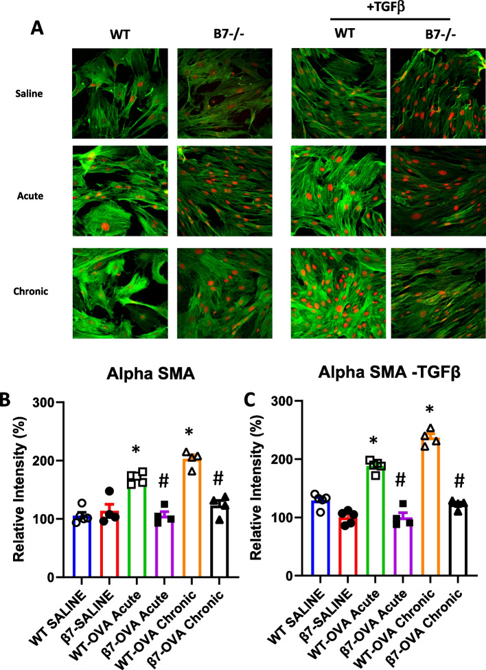
Results: Eosinophil numbers were similar in WT vs β7-/- mice. Prolonged OVA exposure in β7-/- mice was associated with reduced AHR, lung collagen content, peribronchial smooth muscle, lung tissue TGFβ and IL13 expression as compared to WT. Similar findings were observed in WT mice treated with α4β7 blocking antibodies. Fibroblast migration was enhanced in response to OVA in WT but not β7 -/- fibroblasts. α-SMA and fibronectin expression were reduced in β7-/- fibroblasts relative to WT.

Conclusions: The β7 integrin subunit and the α4β7 integrin complex modulate AHR and airway remodeling in a murine model of allergen exposure. This effect is, at least in part, explained by inhibition of fibroblast activation and is independent of eosinophilic inflammation.

Keywords: Airway-hyperresponsiveness; Asthma; Fibroblast; Remodeling; α4β7 integrin.


Conflict of interest statement

The authors declare no competing interests.

Figures

Fig. 1
Fig. 1
Schematic representation of allergen challenge models. Ten-eleven week old C57BL/6 mice were sensitized with intraperitoneal (IP) ovalbumin (OVA) (10 μg OVA/1 mg Al (OH)3 in 0.5 ml 0.9% saline or with saline on days one and 10. Mice were then challenged with inhaled saline or OVA (2% weight/volume diluted in 0.9% saline: 4 ml/inhalation) three times a week for one (“acute”) to 4 weeks (“chronic”), starting on day 15. (A) Allergen challenge using β 7-/- mice or wild-type controls; (B) Mice treated with saline or α4β7 blocking antibody (DAKT32), 100ug in 100µl / dose administered intraperitoneally weekly for 6 weeks
Fig. 2
Fig. 2
Bronchoalveloar cell counts, airway hyperresponsiveness and airway remodeling in β 7-/- and WT mice. A Bronchoalveolar fluid cell counts (per ml) from C57BL/6 wild type (WT) and β 7-/- mice treated with saline or OVA for one ("acute") or four weeks ("chronic"). B Airway hyperresponsiveness (resistance—R) in response to increasing doses of inhaled methacholine (MCH) in wild type and β 7-/- mice treated with saline or OVA for four weeks. Resistance at the maximal MCH dose (64 mg/ml). p = 0.03 for OVA vs SAL in WT mice, p = 0.048 for WT-OVA vs β7 -/- OVA, Area under the curve: p = 0.08 WT-OVA vs β7 -/- OVA. N = 8–10 mice per group. C Soluble collagen content (ug/100 mg tissue) measured by Sircol in the lungs of acute and chronic OVA treated WT and B7-/- mice. D Peribronchial smooth muscle area expressed as pixel number (area/length) using image J Pro Plus software in Masson Trichrome stained lung sections. n = 8–10 mice per group for collagen and 4–5 per group for smooth muscle. E Semi-quantitative score for epithelial goblet cells in PAS-stained sections n = 5 mice per group *p < 0.05 vs WT-SAL and vs β7 -/-SAL, #p < 0.05 vs WT-OVA acute and vs WT-OVA Chronic
Fig. 3
Fig. 3
TGF β and Interleukin-13 expression in β 7-/- and WT mice treated with saline or OVA for one ("acute") or four weeks ("chronic"). A TGF β in BALf, B TGF β in lung tissue C IL-13 in BALf, D IL-13 in lung tissue. *p < 0.05 vs SAL, #p < 0.05 for β7 -/- OVA vs WT-OVA. n = 8–10 mice per group
Fig. 4
Fig. 4
Bronchoalveloar cell counts, airway hyperresponsiveness, collagen and smooth muscle content in the presence or absence of neutralizing anti-α4β7 antibody. C57BL/6 mice were exposed to saline or OVA for 4 weeks in the presence or absence of neutralizing anti-α4β7 antibody (100ug in 100µl PBS given intraperitoneally weekly from day 0, total of 6 doses) or intraperitoneal saline. A Bronchoalveolar fluid cell counts (%). B Airway hyperresponsiveness (Airway / Newtonian resistance—Rn) in response to increasing doses of inhaled methacholine (MCH), p < 0.01 for area under the curve (AUC) and for resistance at the maximal MCH dose (64 mg/ml) for OVA vs OVA + antibody. C lung collagen content, *p < 0.01 for OVA vs SAL, # p = 0.014 for OVA vs OVA + antibody (D, E) Peribronchial smooth muscle in Masson Trichrome stained lung sections: (D) area expressed as pixel number using image J Pro Plus software. E representative histological images, nt: not treated with antibody. *p < 0.05 for OVA vs SAL, # p < 0.05 for OVA vs OVA + antibody. n = 5–8 for each group
Fig. 5
Fig. 5
Lung fibroblast migration from WT and β7-/- mice. A, B Murine lung fibroblasts from WT and from β7-/- mice exposed to saline, OVA one week (acute) or OVA for 4 weeks (chronic exposure) were grown in 12-well culture plates until confluence. Following scratch wound, cell migration is expressed as percentage closure of wound at 16 h. A Representative images (20X and 50X (insert) (B) % recovered wound area. *p < 0.001 WT-OVA vs saline, *p < 0.001 β7-/- OVA vs WT-OVA, n = 4 per group
Fig. 6
Fig. 6
Expression of α-SMA in murine lung fibroblasts from WT and β7-/- mice. Fibroblasts (passage 2) from WT and from β7-/- mice exposed to saline, OVA one week (acute) or OVA for 4 weeks (chronic exposure) were grown in culture plates for 48 h in the presence or absence of TGF-β (10ng/ml). Cells were stained for α-smooth muscle actin (α-SMA) (Sigma Aldrich, USA) (FITC—green) and PI for nuclei (red) (A). Average intensity of fluorescence (expressed as mean % change density/ intensity relative to control mice (WT-Sal) is shown in B (without TGF-β) and C (with TGF-β). N = 4, (* P < 0.05 for WT SAL vs OVA, #p < 0.05 for. β7-/- OVA vs WT-OVA)
Fig. 7
Fig. 7
Expression of fibronectin in murine lung fibroblasts from WT and β7-/- mice. Murine lung fibroblasts (passage 2) from WT and from β7-/- mice exposed to saline, OVA one week (acute) or OVA for 4 weeks (chronic exposure) were grown in culture plates for 48h in the presence or absence of TGF-β (10ng/ml). Cells were stained for total fibronectin (3E2, Abcam, Cambridge, UK) (FITC—green) and PI for nuclei (red) (A). Average intensity of fluorescence (expressed as mean %Change Density/Intensity relative to control mice (WT-SAL) is shown in B (without TGF-β) and C (with TGF-β). N = 4, (* P < 0.05 for WT SAL vs OVA, #p < 0.05 for β7-/- OVA vs WT-OVA

Similar articles

References

    1. Pascual RM, Peters SP. Airway remodeling contributes to the progressive loss of lung function in asthma: an overview. J Allergy Clin Immunol. 2005;116:477–486. doi: 10.1016/j.jaci.2005.07.011. - DOI - PubMed
    1. Jeffery PK. Remodeling in asthma and chronic obstructive lung disease. Am J Respir Crit Care Med. 2001;164:S28–38. doi: 10.1164/ajrccm.164.supplement_2.2106061. - DOI - PubMed
    1. Jeffery PK. Remodeling and Inflammation of bronchi in Asthma and Chronic Obstructive Pulmonary Disease. Proc Am Thorac Soc. 2004;1(3):176–283. doi: 10.1513/pats.200402-009MS. - DOI - PubMed
    1. Fang L. Sun Q, Roth M, Immunologic and non-immunologic mechanisms leading to airway remodeling in asthma. Int J Mol Sci. 2020;21:757. doi: 10.3390/ijms21030757. - DOI - PMC - PubMed
    1. Saglani S, Lloyd CM. Novel Concepts in Airway Inflammation and Remodeling in Asthma. Eur Respir J. 2015;46:1796–1804. doi: 10.1183/13993003.01196-2014. - DOI - PubMed

MeSH terms

LinkOut - more resources