. 2024 Jun 25;14(13):1866.
doi: 10.3390/ani14131866.

Lesion Distribution in the Metacarpophalangeal and Metatarsophalangeal Region of 341 Horses Using Standing Magnetic Resonance Imaging

Affiliations

Lesion Distribution in the Metacarpophalangeal and Metatarsophalangeal Region of 341 Horses Using Standing Magnetic Resonance Imaging

Stefano Schiavo et al. Animals (Basel). .

Abstract

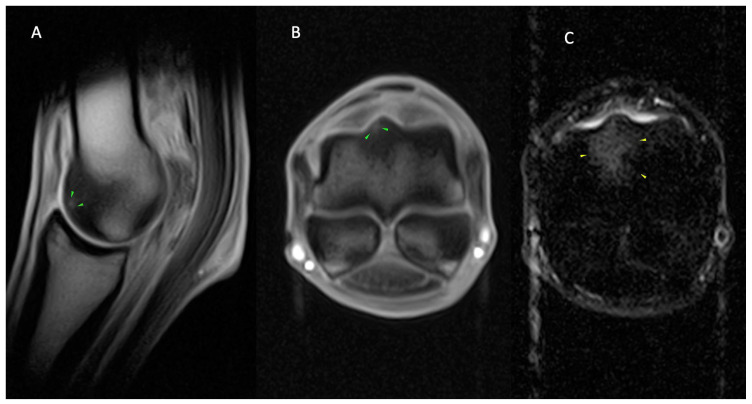
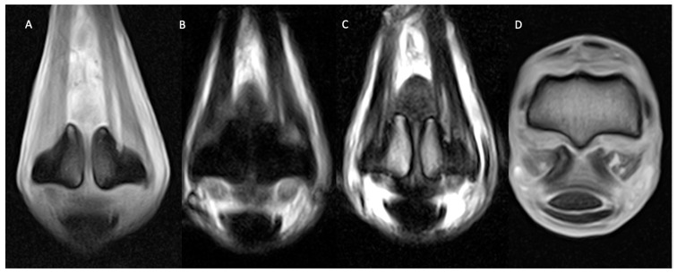
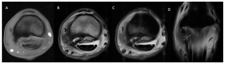
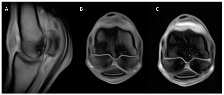
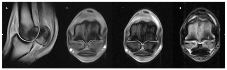
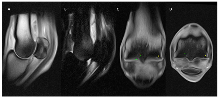
Pain localised to the metacarpophalangeal (MCP) and metatarsophalangeal (MTP) region represents a frequent cause of lameness in sport horses, and standing magnetic resonance imaging (MRI) of these regions is increasingly being acquired. This multicentre retrospective study describes the ranges of abnormalities identified on standing MRI of the MCP/MTP region and compares patterns of abnormalities between forelimbs, hindlimbs and different sports disciplines. In total, 341 MRI reports were reviewed. Subchondral bone plate irregularities, condylar and proximal phalanx pathologies were frequently identified with subchondral bone defect, sclerosis and increased intensity on STIR images often described. Medial pathology was frequently identified in the forelimbs, and more lateral pathology was reported in the hindlimbs, which could potentially reflect differences in the loading patterns. Significant differences in MRI findings were found between different sports activities, with MCP/MTP bone pathology occurring more frequently in MRI reports from race and endurance horses and MCP/MTP soft tissue injuries being reported more frequently in dressage and show-jumping horses, particularly in the suspensory apparatus, including the distal sesamoidean ligaments. The findings of this study identify patterns of MCP/MTP abnormalities detected using standing MRI, with differences seen between forelimbs and hindlimbs and between different sports disciplines.

Keywords: MRI; equine; fetlock; sport horse.


Conflict of interest statement

The authors declare no conflicts of interest.

Figures

Figure 1
Figure 1
Lateral is to the left of the transverse images (B,C). From left, sagittal (A) and transverse T1 GRE (B) and transverse STIR FSE (C) images of the right MCP region of a horse with osseous and joint pathology. Well-defined, focal T1 GRE hyperintensity at the dorsolateral articular margin of the sagittal ridge (green arrows; (A,B)), which is surrounded by T1 GRE hypointense (A,B) and STIR FSE hyperintense signal (yellow arrows; (B)) of the dorsal sagittal ridge and adjacent dorsal half trabecular bone. The findings are consistent with the focal area of subchondral bone injury (green arrows; (A,B)) on the dorsolateral sagittal ridge, surrounded by a larger area of fluid signal ((AC) (yellow arrows)).
Figure 2
Figure 2
Lateral is to the left of the dorsal (AC) and transverse (D) images. From left, dorsal T1 GRE (A), STIR FSE (B), T2 FSE (C), and transverse T1 GRE (D) images of the right MCP region of a horse with soft tissue and osseous pathology. There are well-defined, extensive, longitudinal injuries (hyperintense in all sequences) suggestive of active desmopathy of both suspensory branches (medial > lateral) with mild enlargement of the medial suspensory branch and moderate medial homogeneous periligamentous soft tissue swelling. Moderate abaxial bone remodelling and sclerosis of both proximal sesamoid bones are present with focal, well-defined, triangular-shaped STIR FSE hyperintense area (B) in the bone ligament interface, consistent with active medial enthesopathy.
Figure 3
Figure 3
Lateral is on the left of the transverse (AC) and dorsal (D) images. From left, transverse T1 GRE (A), T2 FSE (B), STIR FSE (C) and dorsal STIR FSE (D) images of the right MCP region of a horse with soft tissue and osseous pathology. Marked enlargement and prominent linear hyperintensity (in all sequences) of the lateral oblique distal sesamoidean ligament. The abnormal ligament is bordered on its lateral aspect by a large T1 GRE hypointense osseous spur (A) and marked T2 FSE heterogeneous periligamentous soft tissue swelling (B) in the region of the lateral aspect of the proximal digital annular ligament and lateral collateral sesamoidean ligament. T1 GRE (A)/T2 FSE (B) hypointense and STIR FSE (C) hyperintense signal in the palmarolateral half trabecular bone of the proximal phalanx. The findings are consistent with marked, active desmopathy and enthesopathy of the lateral oblique distal sesamoidean ligament +/− desmitis of the proximal digital annular ligament and lateral collateral sesamoidean ligament with associated marked bone marrow lesion (oedema-like) in the palmarolateral proximal phalanx.
Figure 4
Figure 4
The lateral is to the right of the transverse images (B,C). From left, sagittal (A) and transverse T1 GRE (B), transverse T2* GRE (C) images of the left MCP region of a horse with a lateral parasagittal groove subchondral bone lesion. A small, focal and ill-defined area of moderate hyperintense signal in all sequences along the palmar aspect of the lateral parasagittal groove (green arrows; (AC)) surrounded by moderate T1 hypointense signal (A,B) and mixed T2* GRE signal intensity with ill-defined fat-water (phase) cancellation artefact (C). Moderate intermediate T1 (A,B) and T2* GRE signal intensity (C) in the dorsal and palmar half of the medial condyle and dorsal half of the sagittal ridge.
Figure 5
Figure 5
Lateral is to the left of the transverse images (AC). From left, sagittal (A) and transverse T1 GRE, (B) transverse T2* GRE (C) and T2 FSE (D) images of the right MCP region of a horse with active bone and joint pathology. Well-defined T1 GRE and T2 FSE hypointensity and T2* GRE fat-water (phase) cancellation artefact is present in the palmar half of the medial third metacarpal condyle with a triangular to the linear area of marked hyperintensity present in all sequences, along the palmarodistal subchondral bone surface of the medial condyle.
Figure 6
Figure 6
Lateral is to the left of the transverse (D) and dorsal (C) images. From left, sagittal T1 GRE (A) and STIR FSE (B), dorsal (C) and transverse T1 GRE (D) images of the right MCP region of a horse with osseous and fissure pathology. Well-defined, short, linear, T1 GRE hyperintensity extending from the palmar aspect of the lateral parasagittal groove (green arrows; (C,D)), surrounded by a large area of mixed T1 GRE hypointense (A,C,D) and STIR FSE hyperintense (B) signal extending throughout both metacarpal condyles. Focal, shallow T1 hyperintense subchondral outline irregularity along the palmar aspect of the medial metacarpal condyle (yellow arrows; (C,D)). The findings are consistent with fissure pathology of the palmar aspect of the lateral parasagittal groove (green arrows; (C,D)) and medial condylar focal area of subchondral bone resorption (yellow arrows; (C,D)) with marked, generalised condylar bone marrow lesion (oedema like) (B) and sclerosis (A,C,D) which are more evident in the lateral condyle.

Similar articles

Cited by

References

    1. Dyson S.J., Murray R.C. Osseous trauma in the fetlock region of mature sports horses; Proceedings of the American Association of Equine Practitioners Annual Convention; San Antonio, TX, USA. 2–6 December 2006; pp. 443–456.
    1. Riggs C., Whitehouse G., Boyde A. Pathology of the distal condyles of the third metacarpal and third metatarsal bones of the horse. Equine Vet. J. 1999;31:140–148. doi: 10.1111/j.2042-3306.1999.tb03807.x. - DOI - PubMed
    1. Riggs C., Whitehouse G., Boyde A. Structural variation of the distal condyle of the third metacarpal and third metatarsal bones in the horse. Equine Vet. J. 1999;31:130–139. doi: 10.1111/j.2042-3306.1999.tb03806.x. - DOI - PubMed
    1. Pool R.R. Multidisciplinary investigation of the aetiopathogenesis of parasagittal fractures of the third metacarpal and metatarsal bones of Thoroughbreds. Equine Vet. J. 1999;31:96. doi: 10.1111/j.2042-3306.1999.tb03800.x. - DOI - PubMed
    1. Gonzalez L.M., Schramme M.C., Robertson I.D., Thrall D.E., Redding R.W. MRI features of metacarpo(tarso)phalangeal region lameness in 40 horses. Vet. Radiol. Ultrasound. 2010;51:404–414. doi: 10.1111/j.1740-8261.2010.01676.x. - DOI - PubMed

LinkOut - more resources