Histological and Histopathological Features of the Third Metacarpal/Tarsal Parasagittal Groove and Proximal Phalanx Sagittal Groove in Thoroughbred Horses with Racing History
- PMID: 38998057
- PMCID: PMC11240324
- DOI: 10.3390/ani14131942
Histological and Histopathological Features of the Third Metacarpal/Tarsal Parasagittal Groove and Proximal Phalanx Sagittal Groove in Thoroughbred Horses with Racing History
Abstract
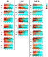
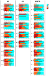
Information regarding the histopathology of the proximal phalanx (P1) sagittal groove in racehorses is limited. Twenty-nine cadaver limbs from nine Thoroughbred racehorses in racing/race-training underwent histological examination. Histological specimens of the third metacarpal/metatarsal (MC3/MT3) parasagittal grooves and P1 sagittal grooves were graded for histopathological findings in hyaline cartilage (HC), calcified cartilage (CC), and subchondral plate and trabecular bone (SCB/TB) regions. Histopathological grades were compared between (1) fissure and non-fissure locations observed in a previous study and (2) dorsal, middle, and palmar/plantar aspects. (1) HC, CC, and SCB/TB grades were more severe in fissure than non-fissure locations in the MC3/MT3 parasagittal groove (p < 0.001). SCB/TB grades were more severe in fissure than non-fissure locations in the P1 sagittal groove (p < 0.001). (2) HC, CC, and SCB/TB grades including SCB collapse were more severe in the palmar/plantar than the middle aspect of the MC3/MT3 parasagittal groove (p < 0.001). SCB/TB grades including SCB collapse were more severe in the dorsal and middle than the palmar/plantar aspect of the P1 sagittal groove (p < 0.001). Histopathology in the SCB/TB region including bone fatigue injury was related to fissure locations, the palmar/plantar MC3/MT3 parasagittal groove, and the dorsal P1 sagittal groove.
Keywords: articular cartilage; fissure; metacarpal/metatarsal parasagittal groove; proximal phalanx sagittal groove; subchondral bone; thoroughbred.
Conflict of interest statement
N. Bolas is employed by Hallmarq Veterinary Imaging.
Figures


Similar articles
-
Prospective, longitudinal assessment of subchondral bone morphology and pathology using standing, cone-beam computed tomography in fetlock joints of 2-year-old Thoroughbred racehorses in their first year of training.Equine Vet J. 2025 Jan;57(1):126-139. doi: 10.1111/evj.14048. Epub 2024 Jan 21. Equine Vet J. 2025. PMID: 38247205
-
Three-Dimensional Imaging and Histopathological Features of Third Metacarpal/Tarsal Parasagittal Groove and Proximal Phalanx Sagittal Groove Fissures in Thoroughbred Horses.Animals (Basel). 2023 Sep 14;13(18):2912. doi: 10.3390/ani13182912. Animals (Basel). 2023. PMID: 37760312 Free PMC article.
-
Biomechanical testing of the calcified metacarpal articular surface and its association with subchondral bone microstructure in Thoroughbred racehorses.Equine Vet J. 2018 Mar;50(2):255-260. doi: 10.1111/evj.12748. Epub 2017 Sep 27. Equine Vet J. 2018. PMID: 28833497
-
Solar angle of the distal phalanx is associated with scintigraphic evidence of subchondral bone injury in the palmar/plantar aspect of the third metacarpal/tarsal condyles in Thoroughbred racehorses.Equine Vet J. 2019 Nov;51(6):720-726. doi: 10.1111/evj.13086. Epub 2019 Mar 19. Equine Vet J. 2019. PMID: 30793363
-
Aetiopathogenesis of parasagittal fractures of the distal condyles of the third metacarpal and third metatarsal bones--review of the literature.Equine Vet J. 1999 Mar;31(2):116-20. doi: 10.1111/j.2042-3306.1999.tb03803.x. Equine Vet J. 1999. PMID: 10213423 Review.
References
Grants and funding
LinkOut - more resources
Full Text Sources

