Review
. 2024 Jun 24;13(13):3667.
doi: 10.3390/jcm13133667.

Neurovascular Issues in Antiphospholipid Syndrome: Arterial Vasculopathy from Small to Large Vessels in a Neuroradiological Perspective

Affiliations
Review

Neurovascular Issues in Antiphospholipid Syndrome: Arterial Vasculopathy from Small to Large Vessels in a Neuroradiological Perspective

Marialuisa Zedde et al. J Clin Med. .

Abstract

Antiphospholipid syndrome (APS) is an autoimmune prothrombotic condition characterized by venous thromboembolism, arterial thrombosis, and pregnancy morbidity. Among neurological manifestations, arterial thrombosis is only one of the possible associated clinical and neuroradiological features. The aim of this review is to address from a neurovascular point of view the multifaceted range of the arterial side of APS. A modern neurovascular approach was proposed, dividing the CNS involvement on the basis of the size of affected arteries, from large to small arteries, and corresponding clinical and neuroradiological issues. Both large-vessel and small-vessel involvement in APS were detailed, highlighting the limitations of the available literature in the attempt to derive some pathomechanisms. APS is a complex disease, and its neurological involvement appears multifaceted and not yet fully characterized, within and outside the diagnostic criteria. The involvement of intracranial large and small vessels appears poorly characterized, and the overlapping with the previously proposed inflammatory manifestations is consistent.

Keywords: APS; MRI; SVD; antiphospholipid syndrome; intracranial occlusion; large vessels; moyamoya; neuroradiology; proliferative vasculopathy; vasculitis.


Conflict of interest statement

The authors declare no conflicts of interest.

Figures
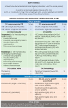
Figure 1
Figure 1
2023 ACR/EULAR APS classification criteria [14]. “Persistent” aPL test results (at least 12 weeks apart) should be scored based on two consecutive positive LAC, two consecutive highest aCL, and/or two consecutive highest aβ2GPI results (two consecutive results with one moderate positive and one high positive aCL/aβ2GPI should be marked as “moderate positive” if there are no additional consecutive high results available). CVD = cardiovascular disease; VTE = venous thromboembolism; AT = arterial thrombosis.
Figure 2
Figure 2
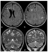
Brain MRI (axial FLAIR in panels (ad), coronal T2W in panels (ag), and axial T2W in panel (h)) showing small punctate white-matter hyperintensities in the centrum semiovale, with a trend to watershed distribution (panel (d)), and a mild increase in enlarged perivascular spaces in the subcortical white matter (panels (eh)).
Figure 3
Figure 3
A remote ischemic lesion in the right MCA territory is illustrated in the axial FLAIR sequence of the brain MRI (panel (ac)) with the corresponding vascular imaging on a CT angiography with minimum intensity projection/multiplanar reconstruction (MIP/MPR) (panel (d,e)) in the coronal and axial plane, respectively. M1 MCA on both sides is occluded with a tiny network of small vessels partially contributing to supply M2 MCA.
Figure 4
Figure 4
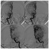
Digital subtraction angiography (DSA) of the same patient as in Figure 3 from a (right) (panels (a,b)) and (left) (panels (c,d)) ICA injection in PA view. Panels (a,c) are an early arterial phase, and panels (a,b) are a mid– late arterial phase. On both sides, the M1 MCA after its origin appears steno-occluded and is substituted by a network of collateral vessels involving the perforating arteries.
Figure 5
Figure 5
An example of multifocal cerebellar ischemic lesions in a patient with APS (double positivity). Brain MRI (axial FLAIR in (a,b), coronal T2W sequence in (c) shows the poromalacic evolution of multiple ischemic lesions involving both cerebellar hemispheres (right ≥ left). No causes other than APS were identified in this patients.
Figure 6
Figure 6
Brain MRI (axial FLAIR in panels (ac) and the corresponding DWI slices in panels (df)) showing multifocal ischemic lesions in the left PCA territory with a patent PCA on MRA (panel (g)).
Figure 7
Figure 7
Brain MRI (axial FLAIR in panels (a,b) and coronal T2W sequence in panels (c,d)) showing multiple cortico-subcortical ischemic lesions on both hemispheres and only few mildest SVD markers.
Figure 8
Figure 8
Brain MRI (axial FLAIR) at baseline (panels (ac)) and after 8 years (panels (d-f)), showing the increase of WMHs in the subcortical white matter.

References

    1. Hughes G.R. The antiphospholipid syndrome: Ten years on. Lancet. 1993;342:341–344. doi: 10.1016/0140-6736(93)91477-4. - DOI - PubMed
    1. Corban M.T., Duarte-Garcia A., McBane R.D., Matteson E.L., Lerman L.O., Lerman A. Antiphospholipid Syndrome: Role of Vascular Endothelial Cells and Implications for Risk Stratification and Targeted Therapeutics. J. Am. Coll. Cardiol. 2017;69:2317–2330. doi: 10.1016/j.jacc.2017.02.058. - DOI - PubMed
    1. Cervera R. Antiphospholipid syndrome. Thromb. Res. 2017;151((Suppl. S1)):S43–S47. doi: 10.1016/S0049-3848(17)30066-X. - DOI - PubMed
    1. Schreiber K., Sciascia S., de Groot P.G., Devreese K., Jacobsen S., Ruiz-Irastorza G., Salmon J.E., Shoenfeld Y., Shovman O., Hunt B.J. Antiphospholipid syndrome. Nat. Rev. Dis. Prim. 2018;4:17103. doi: 10.1038/nrdp.2017.103. - DOI - PubMed
    1. Vila P., Hernández M.C., López-Fernández M.F., Batlle J. Prevalence, follow-up and clinical significance of the anticardiolipin antibodies in normal subjects. Thromb. Haemost. 1994;72:209–213. doi: 10.1055/s-0038-1648840. - DOI - PubMed

LinkOut - more resources