

Developing the Benchmark: Establishing a Gold Standard for the Evaluation of AI Caries Diagnostics
- PMID: 38999411
- PMCID: PMC11242122
- DOI: 10.3390/jcm13133846
Developing the Benchmark: Establishing a Gold Standard for the Evaluation of AI Caries Diagnostics
Abstract
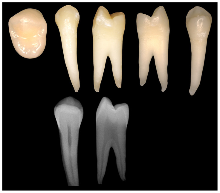
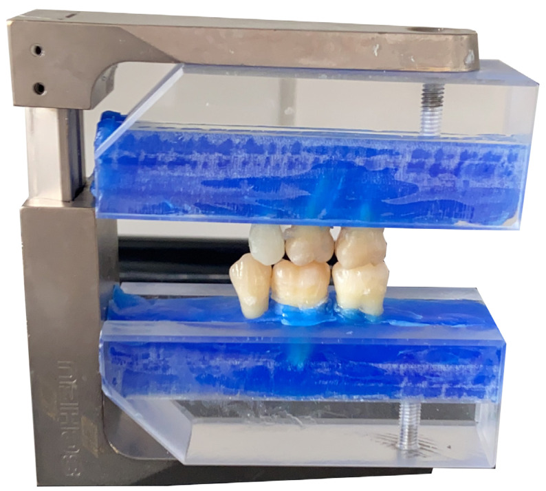
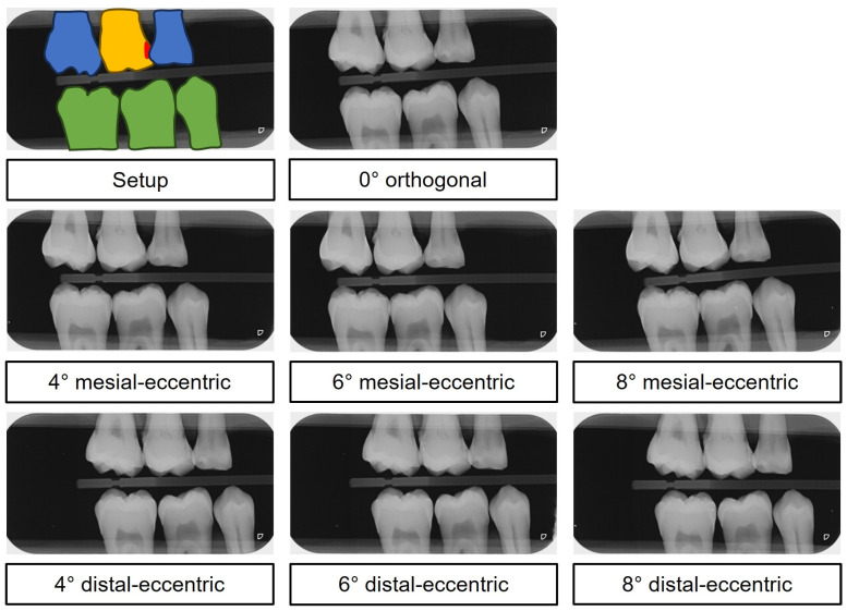
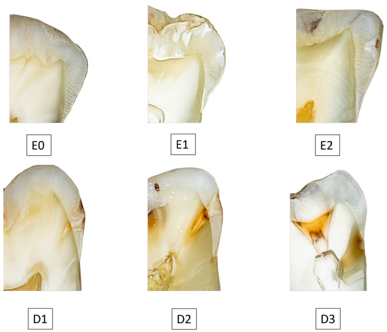
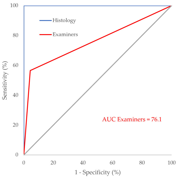
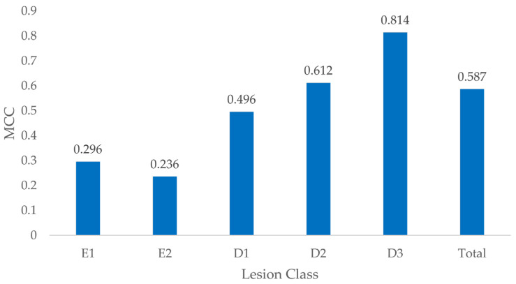
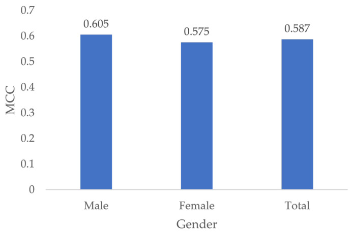
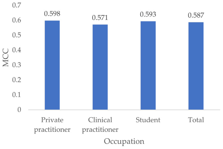
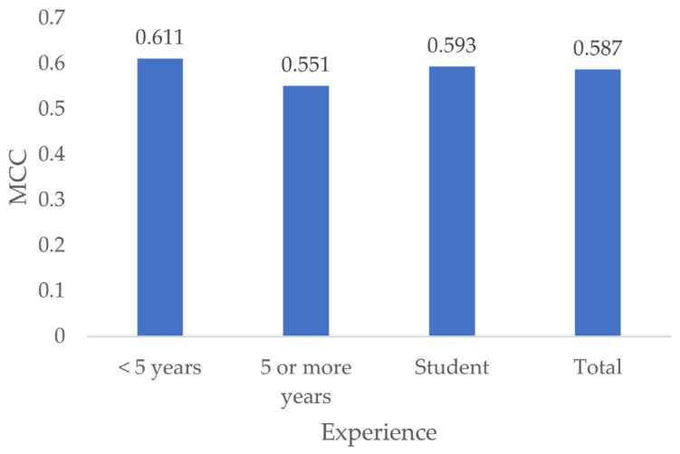
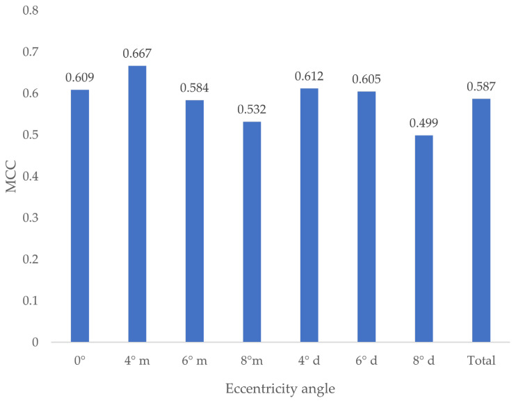
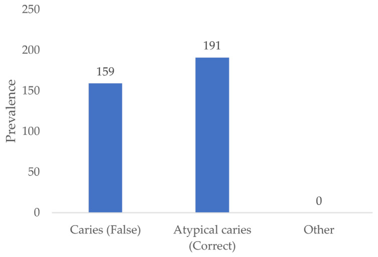
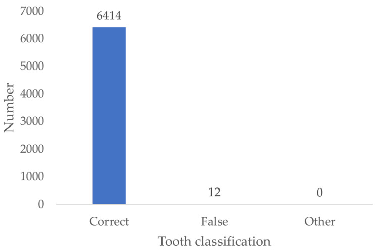
Background/Objectives: The aim of this study was to establish a histology-based gold standard for the evaluation of artificial intelligence (AI)-based caries detection systems on proximal surfaces in bitewing images. Methods: Extracted human teeth were used to simulate intraoral situations, including caries-free teeth, teeth with artificially created defects and teeth with natural proximal caries. All 153 simulations were radiographed from seven angles, resulting in 1071 in vitro bitewing images. Histological examination of the carious lesion depth was performed twice by an expert. A total of thirty examiners analyzed all the radiographs for caries. Results: We generated in vitro bitewing images to evaluate the performance of AI-based carious lesion detection against a histological gold standard. All examiners achieved a sensitivity of 0.565, a Matthews correlation coefficient (MCC) of 0.578 and an area under the curve (AUC) of 76.1. The histology receiver operating characteristic (ROC) curve significantly outperformed the examiners' ROC curve (p < 0.001). All examiners distinguished induced defects from true caries in 54.6% of cases and correctly classified 99.8% of all teeth. Expert caries classification of the histological images showed a high level of agreement (intraclass correlation coefficient (ICC) = 0.993). Examiner performance varied with caries depth (p ≤ 0.008), except between E2 and E1 lesions (p = 1), while central beam eccentricity, gender, occupation and experience had no significant influence (all p ≥ 0.411). Conclusions: This study successfully established an unbiased dataset to evaluate AI-based caries detection on bitewing surfaces and compare it to human judgement, providing a standardized assessment for fair comparison between AI technologies and helping dental professionals to select reliable diagnostic tools.
Keywords: artificial intelligence; benchmarking; bitewing; dental caries; diagnostics; radiography.
Conflict of interest statement
The authors declare no potential conflicts of interest concerning the research, authorship and/or publication of this article.
Figures

Similar articles
-
Accuracy of Digital Bitewing Radiography versus Different Views of Digital Panoramic Radiography for Detection of Proximal Caries.J Dent (Tehran). 2015 Apr;12(4):290-7. J Dent (Tehran). 2015. PMID: 26622284 Free PMC article.
-
Comparison of proximal caries detection in primary teeth between laser fluorescence and bitewing radiography.Pediatr Dent. 2005 Nov-Dec;27(6):493-9. Pediatr Dent. 2005. PMID: 16532891
-
Intraoral versus extraoral bitewing radiography in detection of enamel proximal caries: an ex vivo study.Dentomaxillofac Radiol. 2016;45(4):20150326. doi: 10.1259/dmfr.20150326. Epub 2016 Feb 19. Dentomaxillofac Radiol. 2016. PMID: 26892946 Free PMC article.
-
Proximal caries detection accuracy using intraoral bitewing radiography, extraoral bitewing radiography and panoramic radiography.Dentomaxillofac Radiol. 2012 Sep;41(6):450-9. doi: 10.1259/dmfr/30526171. Dentomaxillofac Radiol. 2012. PMID: 22868296 Free PMC article.
-
Diagnostic performance of artificial intelligence-aided caries detection on bitewing radiographs: a systematic review and meta-analysis.Jpn Dent Sci Rev. 2024 Dec;60:128-136. doi: 10.1016/j.jdsr.2024.02.001. Epub 2024 Feb 29. Jpn Dent Sci Rev. 2024. PMID: 38450159 Free PMC article. Review.
Cited by
-
AI in Medical Imaging and Image Processing.J Clin Med. 2025 Jun 11;14(12):4153. doi: 10.3390/jcm14124153. J Clin Med. 2025. PMID: 40565899 Free PMC article.
-
Integrating CT radiomics and clinical data with machine learning to predict fibrosis progression in coalworker pneumoconiosis.Front Med (Lausanne). 2025 Jul 22;12:1599739. doi: 10.3389/fmed.2025.1599739. eCollection 2025. Front Med (Lausanne). 2025. PMID: 40766061 Free PMC article.
References
LinkOut - more resources
Full Text Sources
