Immunoexpression Pattern of Autophagy-Related Proteins in Human Congenital Anomalies of the Kidney and Urinary Tract
- PMID: 38999938
- PMCID: PMC11241479
- DOI: 10.3390/ijms25136829
Immunoexpression Pattern of Autophagy-Related Proteins in Human Congenital Anomalies of the Kidney and Urinary Tract
Abstract
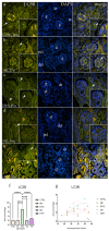
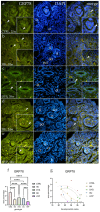
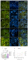
The purpose of this study was to evaluate the spatiotemporal immunoexpression pattern of microtubule-associated protein 1 light chain 3 beta (LC3B), glucose-regulated protein 78 (GRP78), heat shock protein 70 (HSP70), and lysosomal-associated membrane protein 2A (LAMP2A) in normal human fetal kidney development (CTRL) and kidneys affected with congenital anomalies of the kidney and urinary tract (CAKUT). Human fetal kidneys (control, horseshoe, dysplastic, duplex, and hypoplastic) from the 18th to the 38th developmental week underwent epifluorescence microscopy analysis after being stained with antibodies. Immunoreactivity was quantified in various kidney structures, and expression dynamics were examined using linear and nonlinear regression modeling. The punctate expression of LC3B was observed mainly in tubules and glomerular cells, with dysplastic kidneys displaying distinct staining patterns. In the control group's glomeruli, LAMP2A showed a sporadic, punctate signal; in contrast to other phenotypes, duplex kidneys showed significantly stronger expression in convoluted tubules. GRP78 had a weaker expression in CAKUT kidneys, especially hypoplastic ones, while normal kidneys exhibited punctate staining of convoluted tubules and glomeruli. HSP70 staining varied among phenotypes, with dysplastic and hypoplastic kidneys exhibiting stronger staining compared to controls. Expression dynamics varied among observed autophagy markers and phenotypes, indicating their potential roles in normal and dysfunctional kidney development.
Keywords: CAKUT; GRP78; HSP70; LAMP2A; LC3B; autophagy; congenital anomalies of the kidney and urinary tract; nephrogenesis.
Conflict of interest statement
The authors declare no conflicts of interest. The funders had no role in the design of the study, nor in the collection, analyses, or interpretation of data, in the writing of the manuscript, or in the decision to publish the results.
Figures


Similar articles
-
Expression of Autophagy Markers LC3B, LAMP2A, and GRP78 in the Human Kidney during Embryonic, Early Fetal, and Postnatal Development and Their Significance in Diabetic Kidney Disease.Int J Mol Sci. 2024 Aug 23;25(17):9152. doi: 10.3390/ijms25179152. Int J Mol Sci. 2024. PMID: 39273100 Free PMC article.
-
Spatiotemporal distribution of Wnt signaling pathway markers in human congenital anomalies of kidney and urinary tract.Acta Histochem. 2025 Mar;127(1):152235. doi: 10.1016/j.acthis.2025.152235. Epub 2025 Feb 4. Acta Histochem. 2025. PMID: 39908631
-
Expression Profiles of ITGA8 and VANGL2 Are Altered in Congenital Anomalies of the Kidney and Urinary Tract (CAKUT).Molecules. 2024 Jul 12;29(14):3294. doi: 10.3390/molecules29143294. Molecules. 2024. PMID: 39064873 Free PMC article.
-
Anatomy and embryology of congenital surgical anomalies: Congenital Anomalies of the Kidney and Urinary Tract.Semin Pediatr Surg. 2022 Dec;31(6):151232. doi: 10.1016/j.sempedsurg.2022.151232. Epub 2022 Nov 17. Semin Pediatr Surg. 2022. PMID: 36423515 Review.
-
Genetics of congenital anomalies of the kidney and urinary tract.Pediatr Nephrol. 2011 Mar;26(3):353-64. doi: 10.1007/s00467-010-1629-4. Epub 2010 Aug 27. Pediatr Nephrol. 2011. PMID: 20798957 Review.
Cited by
-
Mitochondrial Dysfunction: The Silent Catalyst of Kidney Disease Progression.Cells. 2025 May 28;14(11):794. doi: 10.3390/cells14110794. Cells. 2025. PMID: 40497970 Free PMC article. Review.
-
Expression of Autophagy Markers LC3B, LAMP2A, and GRP78 in the Human Kidney during Embryonic, Early Fetal, and Postnatal Development and Their Significance in Diabetic Kidney Disease.Int J Mol Sci. 2024 Aug 23;25(17):9152. doi: 10.3390/ijms25179152. Int J Mol Sci. 2024. PMID: 39273100 Free PMC article.
-
Analysis of Kallikrein 6, Acetyl-α-Tubulin, and Aquaporin 1 and 2 Expression Patterns During Normal Human Nephrogenesis and in Congenital Anomalies of the Kidney and Urinary Tract (CAKUT).Genes (Basel). 2025 Apr 27;16(5):499. doi: 10.3390/genes16050499. Genes (Basel). 2025. PMID: 40428321 Free PMC article.
References
-
- Saran R., Robinson B., Abbott K.C., Bragg-Gresham J., Chen X., Gipson D., Gu H., Hirth R.A., Hutton D., Jin Y., et al. US Renal Data System 2019 Annual Data Report: Epidemiology of Kidney Disease in the United States. Am. J. Kidney Dis. Off. J. Natl. Kidney Found. 2020;75:A6–A7. doi: 10.1053/j.ajkd.2019.09.003. - DOI - PubMed
-
- Xie Y., Bowe B., Mokdad A.H., Xian H., Yan Y., Li T., Maddukuri G., Tsai C.Y., Floyd T., Al-Aly Z. Analysis of the Global Burden of Disease study highlights the global, regional, and national trends of chronic kidney disease epidemiology from 1990 to 2016. Kidney Int. 2018;94:567–581. doi: 10.1016/j.kint.2018.04.011. - DOI - PubMed
MeSH terms
Substances
Supplementary concepts
Grants and funding
LinkOut - more resources
Full Text Sources
Research Materials
Miscellaneous

