. 2024 Jun 22;25(13):6872.
doi: 10.3390/ijms25136872.

LPA-Induced Thromboxane A2-Mediated Vasoconstriction Is Limited to Poly-Unsaturated Molecular Species in Mouse Aortas

Affiliations

LPA-Induced Thromboxane A2-Mediated Vasoconstriction Is Limited to Poly-Unsaturated Molecular Species in Mouse Aortas

Krisztina Vén et al. Int J Mol Sci. .

Abstract

We have previously reported that, in aortic rings, 18:1 lysophosphatidic acid (LPA) can induce both vasodilation and vasoconstriction depending on the integrity of the endothelium. The predominant molecular species generated in blood serum are poly-unsaturated LPA species, yet the vascular effects of these species are largely unexplored. We aimed to compare the vasoactive effects of seven naturally occurring LPA species in order to elucidate their potential pathophysiological role in vasculopathies. Vascular tone was measured using myography, and thromboxane A2 (TXA2) release was detected by ELISA in C57Bl/6 mouse aortas. The Ca2+-responses to LPA-stimulated primary isolated endothelial cells were measured by Fluo-4 AM imaging. Our results indicate that saturated molecular species of LPA elicit no significant effect on the vascular tone of the aorta. In contrast, all 18 unsaturated carbon-containing (C18) LPAs (18:1, 18:2, 18:3) were effective, with 18:1 LPA being the most potent. However, following inhibition of cyclooxygenase (COX), these LPAs induced similar vasorelaxation, primarily indicating that the vasoconstrictor potency differed among these species. Indeed, C18 LPA evoked a similar Ca2+-signal in endothelial cells, whereas in endothelium-denuded aortas, the constrictor activity increased with the level of unsaturation, correlating with TXA2 release in intact aortas. COX inhibition abolished TXA2 release, and the C18 LPA induced vasoconstriction. In conclusion, polyunsaturated LPA have markedly increased TXA2-releasing and vasoconstrictor capacity, implying potential pathophysiological consequences in vasculopathies.

Keywords: lysophosphatidic acid; lysophosphatidic acid receptor 1; thromboxane; vasoconstriction.


Conflict of interest statement

The authors declare no conflicts of interest.

Figures

Figure 1
Figure 1
Vasorelaxation evoked by saturated molecular species of LPA. (a) Representative recordings of vasorelaxation induced by saturated LPA in PE precontracted thoracic aorta segments prepared from WT mice. Arrows indicate the administration of 10 µM LPA; W, here and in subsequent figures, denotes the washout. (b) Vasorelaxation induced by 10 µM 14:0 and 16:0 LPA in aortic rings prepared from WT and Lpar1 KO mice (n = 6, 6 WT and 5, 4 LPA1 KO; two-way ANOVA; ** p < 0.01 vs. WT; *** p < 0.001 vs. WT ). (c) Vasorelaxation induced by 10 µM 18:0 and 20:0 LPA in aortic segments isolated from WT mice (n = 5, 4 unpaired Student’s t-test). Bars represent mean ± SEM.
Figure 2
Figure 2
Vasoconstriction evoked by saturated molecular species of LPA. (a) Representative recordings of vasoconstriction induced by saturated LPA in endothelium-denuded aortic segments prepared from WT mice. Arrows indicate the administration of 10 µM LPA. (b) Vasoconstriction induced by 10 µM 14:0 and 16:0 LPA in endothelium-denuded aortic rings isolated from WT and Lpar1 KO mice (n = 6, 9 for WT and 4, 5 for LPA1 KO; two-way ANOVA; ** p < 0.01 vs. WT). (c) Vasoconstriction induced by 10 µM 18:0 and 20:0 LPA in endothelium-denuded aortic rings isolated from WT mice (n = 5, 3; unpaired Student’s t-test). Bars represent mean ± SEM.
Figure 3
Figure 3
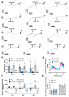
Vasorelaxation induced by unsaturated molecular species of LPA. (a) Representative recordings of vasorelaxation elicited by unsaturated LPA in PE precontracted aortic rings isolated from WT mice. Arrows indicate the administration of 10 µM LPA. (b) Representative recordings of vasorelaxation elicited by 10 µM of unsaturated LPA in PE-precontracted aortic segments isolated from Lpar1 knockout (KO) mice. (c) Representative recordings of vasorelaxation elicited by 10 µM of unsaturated LPA in PE-precontracted aortic segments treated with INDO. (d) Representative recordings of vasorelaxation elicited by 10 µM of unsaturated LPA in PE-precontracted aortic segments isolated from eNOS KO mice. (e) Maximal relaxation activity elicited by 10 µM 18:1, 18:2, and 18:3 LPA in aortic rings isolated from WT, Lpar1 KO, eNOS KO mice, or WT aortic segments treated with INDO (WT + INDO) [n = 18, 21, 25 (WT); 5, 8, 10 (LPA1 KO); 14, 18, 14 (WT + INDO); 4, 4, 4 (eNOS KO) two-way ANOVA; *** p < 0.001 vs. 18:1 WT, ‡ p < 0.05 vs. 18:2 WT, ## p < 0.01 vs. own WT, #### p < 0.0001 vs. own WT]. (f) Area under the vascular response curve in the first 5 min induced by C18 LPA (10 µM of each) in aortic rings isolated from WT, Lpar1 KO, eNOS KO mice, or WT aortic segments treated with INDO (WT + INDO) [n = 15, 21, 25 (WT); 5, 8, 10 (LPA1 KO); 9, 18, 14 (WT + INDO); n = 4, 4, 4 (eNOS KO); two-way ANOVA; *** p < 0.001 vs. 18:1 WT, **** p < 0.0001 vs. 18:1 WT, ‡ p < 0.05 vs. 18:2 WT, # p < 0.05 vs. own WT, ### p < 0.001 vs. own WT, #### p < 0.0001 vs. own WT]. Positive bars indicate dominant relaxation, whereas negative bars indicate a dominant constrictor response. Calculation method for the “area of vascular response” is shown in Supplementary Materials. (g) Average trace of fluorescent intensity in Fluo-4 AM-loaded endothelial cells isolated from the aortas of WT mice. Administration of 10 µM of C18 LPA and 10 µM of ATP indicated by arrows (n = 4, 4, 3). (h) Maximal increase in fluorescent intensity evoked by either 10 µM C18 LPA or ATP (n = 4, 4, 3 for LPA; 4, 4, 3 for ATP; one-way ANOVA). Bars represent mean ± SEM.
Figure 4
Figure 4
Vasoconstriction evoked by unsaturated molecular species of LPA. (a) Representative recordings of vasoconstriction induced by 10 µM of unsaturated LPA in endothelium-denuded AA rings prepared from WT mice. Arrows indicate the administration of 10 µM LPA. (b) Dose–response curves for 18:1, 18:2, and 18:3 LPA-induced vasoconstriction in endothelium-denuded AA rings isolated from WT mice (n = 3–25; two-way ANOVA; ** p < 0.01 vs. 18:1 LPA, **** p < 0.0001 vs. 18:1 LPA, #### p < 0.0001 vs. 18:2 LPA). Bars represent mean ± SEM.
Figure 5
Figure 5
Signaling mechanisms underlying the constrictor activity of unsaturated LPA species. (a) Vasoconstriction elicited by LPA species 18:1, 18:2, and 18:3 (10 µM of each) in endothelium-denuded AA segments isolated from WT and Lpar1 KO mice [n = 9, 10, 11 for WT and 6, 9, 10 for LPA1 KO; two-way ANOVA; *** p < 0.001 vs. 18:1 WT, **** p < 0.0001 vs. 18:1 WT, ‡‡ p < 0.01 vs. 18:2 WT, #### p < 0.0001 vs. own WT]. (b) Vasoconstriction induced by C18 LPA (10 µM of each) in control (Vehicle) and cyclooxygenase-inhibited (INDO) endothelium-denuded vessels isolated from WT mice [n = 4, 7, 6 for vehicle and 4, 7, 7 for Indo; two-way ANOVA; *** p < 0.001 vs. 18:1 WT, **** p < 0.0001 vs. 18:1 WT, #### p < 0.0001 vs. own WT]. (c) TXB2 production in aortas before (Control) and after (Activated) treatment with 10 µM of C18 LPA (n = 4, 4, 3 for control and 4, 4, 3 for activated vessels; two-way ANOVA; * p < 0.05 vs. 18:1 Control, *** p < 0.001 vs. 18:1 control, ## p < 0.01 vs. own control] (d) Vasoconstriction elicited by C18 LPA (10 µM of LPA 18:1, 18:2 and 18:3) in endothelium-denuded aortic rings isolated from mice treated with vehicle (WT) or pertussis toxin (PTX-treated)) [n = 6, 4, 6 for WT and 6, 7, 6 for PTX treated; two-way ANOVA; ** p < 0.01 vs. 18:1 WT, *** p < 0.001 vs. 18:1 WT, # p < 0.05 vs. own WT, ## p < 0.01 vs. own WT]. Bars represent mean ± SEM.
Figure 6
Figure 6
Vasorelaxation and vasoconstriction evoked by a combination of LPA species with a mixture resembling that of plasma. (a) Representative recordings of the vascular effect induced by 100 nM (top) and 10 µM (bottom) of the combination of LPA species in intact TA rings prepared from WT mice. Arrows indicate the administration of the combination; W stands for wash out. (b) Evaluation of the amplitude of vasorelaxation of the LPA mixture [n = 5 for 100 nM, 7 for 10 µM]. (c) Area of vascular response of the LPA mixture [n = 5 for 100 nM, 7 for 10 µM; unpaired t-test, * p < 0.05 vs. 100 nM]. (d) Representative recordings of vasoconstriction induced by 100 nm (top) and 10 µM (bottom) of the combination of LPA species in endothelium-denuded AA rings prepared from WT mice. Arrows indicate the administration of the combination; W stands for washing out. (e) Vasoconstriction induced by 100 nM and 10 µM of the combination of LPA species in endothelium-denuded AA rings isolated from WT mice [n = 4 for 100 nM, 6 for 10 µM; unpaired t-test, ** p < 0.01 vs. 100 nM]. Bars represent mean ± SEM. (f) Relative abundance of the individual LPA molecular species, represented in a pie chart.
Figure 7
Figure 7
Proposed role of naturally occurring LPA species in vascular health and disease.

Similar articles

Cited by

References

    1. Gonzalez-Gil I., Zian D., Vazquez-Villa H., Hernandez-Torres G., Martinez R.F., Khiar-Fernandez N., Rivera R., Kihara Y., Devesa I., Mathivanan S., et al. A Novel Agonist of the Type 1 Lysophosphatidic Acid Receptor (LPA(1)), UCM-05194, Shows Efficacy in Neuropathic Pain Amelioration. J. Med. Chem. 2020;63:2372–2390. doi: 10.1021/acs.jmedchem.9b01287. - DOI - PMC - PubMed
    1. Axelsson Raja A., Wakimoto H., DeLaughter D.M., Reichart D., Gorham J., Conner D.A., Lun M., Probst C.K., Sakai N., Knipe R.S., et al. Ablation of lysophosphatidic acid receptor 1 attenuates hypertrophic cardiomyopathy in a mouse model. Proc. Natl. Acad. Sci. USA. 2022;119:e2204174119. doi: 10.1073/pnas.2204174119. - DOI - PMC - PubMed
    1. Birgbauer E. Lysophosphatidic Acid Signalling in Nervous System Development and Function. Neuromolecular Med. 2021;23:68–85. doi: 10.1007/s12017-020-08630-2. - DOI - PMC - PubMed
    1. Torres R.M., Turner J.A., D’Antonio M., Pelanda R., Kremer K.N. Regulation of CD8 T-cell signaling, metabolism, and cytotoxic activity by extracellular lysophosphatidic acid. Immunol. Rev. 2023;317:203–222. doi: 10.1111/imr.13208. - DOI - PMC - PubMed
    1. Cheng Y., Makarova N., Tsukahara R., Guo H., Shuyu E., Farrar P., Balazs L., Zhang C., Tigyi G. Lysophosphatidic acid-induced arterial wall remodeling: Requirement of PPARgamma but not LPA1 or LPA2 GPCR. Cell. Signal. 2009;21:1874–1884. doi: 10.1016/j.cellsig.2009.08.003. - DOI - PMC - PubMed

LinkOut - more resources