

G Protein-Coupled Receptor-Ligand Pose and Functional Class Prediction
- PMID: 38999982
- PMCID: PMC11241240
- DOI: 10.3390/ijms25136876
G Protein-Coupled Receptor-Ligand Pose and Functional Class Prediction
Abstract
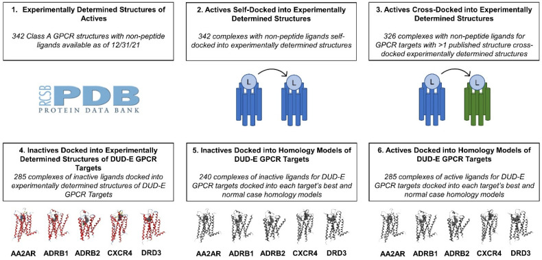
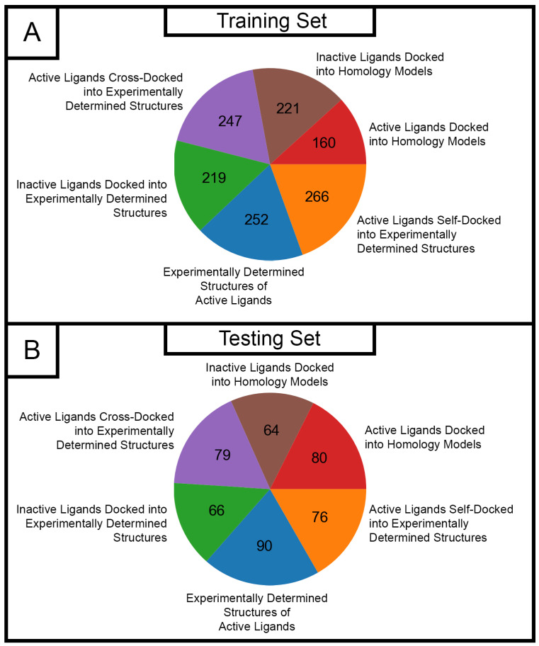
G protein-coupled receptor (GPCR) transmembrane protein family members play essential roles in physiology. Numerous pharmaceuticals target GPCRs, and many drug discovery programs utilize virtual screening (VS) against GPCR targets. Improvements in the accuracy of predicting new molecules that bind to and either activate or inhibit GPCR function would accelerate such drug discovery programs. This work addresses two significant research questions. First, do ligand interaction fingerprints provide a substantial advantage over automated methods of binding site selection for classical docking? Second, can the functional status of prospective screening candidates be predicted from ligand interaction fingerprints using a random forest classifier? Ligand interaction fingerprints were found to offer modest advantages in sampling accurate poses, but no substantial advantage in the final set of top-ranked poses after scoring, and, thus, were not used in the generation of the ligand-receptor complexes used to train and test the random forest classifier. A binary classifier which treated agonists, antagonists, and inverse agonists as active and all other ligands as inactive proved highly effective in ligand function prediction in an external test set of GPR31 and TAAR2 candidate ligands with a hit rate of 82.6% actual actives within the set of predicted actives.
Keywords: G protein-coupled receptor (GPCR); docking; interaction fingerprint; machine learning; random forest classifier.
Conflict of interest statement
The authors declare no conflicts of interest.
Figures
Similar articles
-
Molecular interaction fingerprint approaches for GPCR drug discovery.Curr Opin Pharmacol. 2016 Oct;30:59-68. doi: 10.1016/j.coph.2016.07.007. Epub 2016 Jul 29. Curr Opin Pharmacol. 2016. PMID: 27479316 Review.
-
Self-docking and cross-docking simulations of G protein-coupled receptor-ligand complexes: Impact of ligand type and receptor activation state.J Mol Graph Model. 2022 May;112:108119. doi: 10.1016/j.jmgm.2021.108119. Epub 2021 Dec 28. J Mol Graph Model. 2022. PMID: 34979368
-
Virtual Screening of Human Class-A GPCRs Using Ligand Profiles Built on Multiple Ligand-Receptor Interactions.J Mol Biol. 2020 Aug 7;432(17):4872-4890. doi: 10.1016/j.jmb.2020.07.003. Epub 2020 Jul 9. J Mol Biol. 2020. PMID: 32652079 Free PMC article.
-
Structure-Based Prediction of G-Protein-Coupled Receptor Ligand Function: A β-Adrenoceptor Case Study.J Chem Inf Model. 2015 May 26;55(5):1045-61. doi: 10.1021/acs.jcim.5b00066. Epub 2015 May 1. J Chem Inf Model. 2015. PMID: 25848966
-
Approaches for Differentiation and Interconverting GPCR Agonists and Antagonists.Methods Mol Biol. 2018;1705:265-296. doi: 10.1007/978-1-4939-7465-8_12. Methods Mol Biol. 2018. PMID: 29188567 Review.
References
-
- So S.S., Ngo T., Keov P., Smith N.J., Kufareva I. GPCRs. Elsevier; Amsterdam, The Netherlands: 2020. Tackling the Complexities of Orphan GPCR Ligand Discovery with Rationally Assisted Approaches; pp. 295–334.
MeSH terms
Substances
LinkOut - more resources
Full Text Sources
