. 2024 Jun 22;25(13):6873.
doi: 10.3390/ijms25136873.

Functional and Structural Properties of Cytoplasmic Tropomyosin Isoforms Tpm1.8 and Tpm1.9

Affiliations

Functional and Structural Properties of Cytoplasmic Tropomyosin Isoforms Tpm1.8 and Tpm1.9

Ksenia K Lapshina et al. Int J Mol Sci. .

Abstract

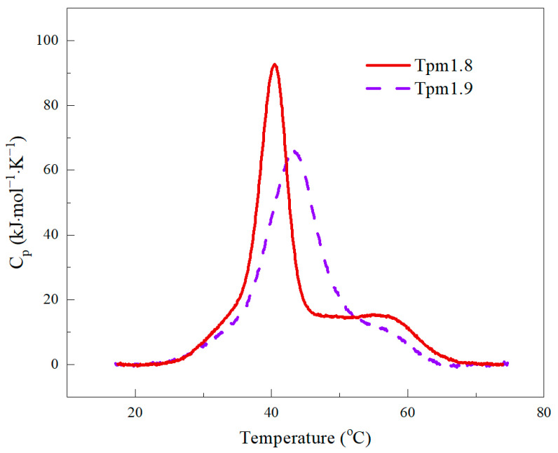
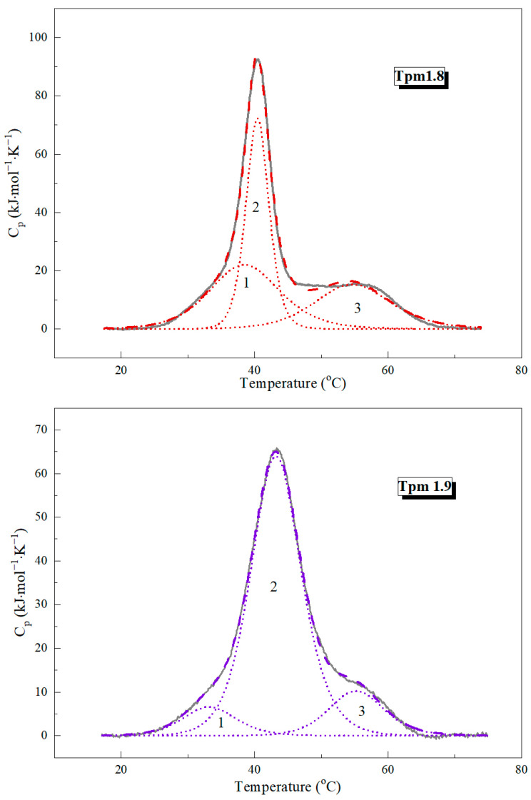
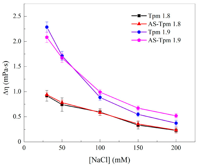
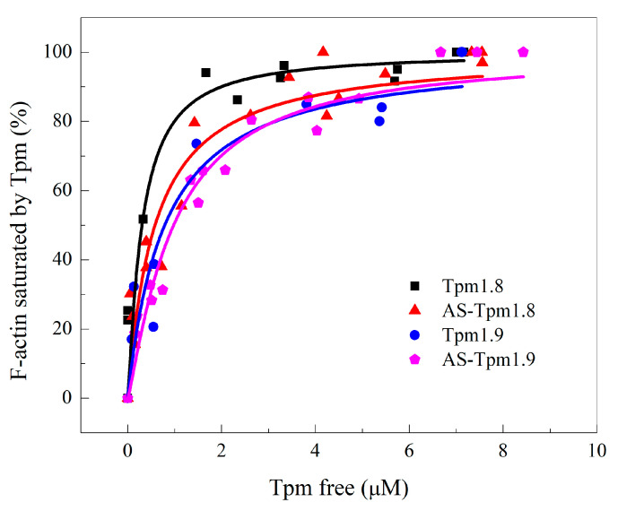
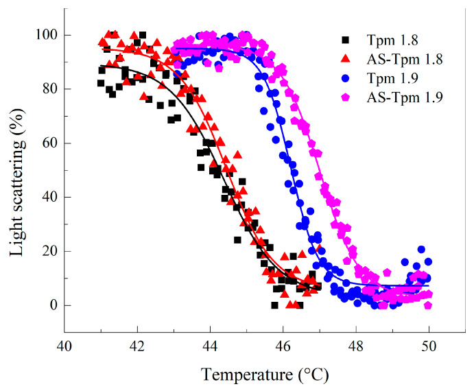
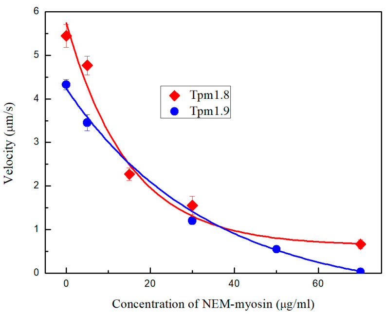
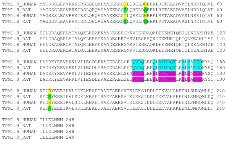
The actin cytoskeleton is one of the most important players in cell motility, adhesion, division, and functioning. The regulation of specific microfilament formation largely determines cellular functions. The main actin-binding protein in animal cells is tropomyosin (Tpm). The unique structural and functional diversity of microfilaments is achieved through the diversity of Tpm isoforms. In our work, we studied the properties of the cytoplasmic isoforms Tpm1.8 and Tpm1.9. The results showed that these isoforms are highly thermostable and differ in the stability of their central and C-terminal fragments. The properties of these isoforms were largely determined by the 6th exons. Thus, the strength of the end-to-end interactions, as well as the affinity of the Tpm molecule for F-actin, differed between the Tpm1.8 and Tpm1.9 isoforms. They were determined by whether an alternative internal exon, 6a or 6b, was included in the Tpm isoform structure. The strong interactions of the Tpm1.8 and Tpm1.9 isoforms with F-actin led to the formation of rigid actin filaments, the stiffness of which was measured using an optical trap. It is quite possible that the structural and functional features of the Tpm isoforms largely determine the appearance of these isoforms in the rigid actin structures of the cell cortex.

Keywords: actin cytoskeleton dynamics; actin filaments; actin-associated proteins; cytoplasmic isoforms of tropomyosin; differential scanning calorimetry; optical trap.


Conflict of interest statement

The authors declare no conflicts of interest.

Figures

Figure 1
Figure 1
The DSC profiles of the excess heat capacity obtained for Tpm1.8 and Tpm1.9.
Figure 2
Figure 2
The deconvolution analysis of the heat absorption curves of Tpm1.8 and Tpm1.9. The solid curves represent the experimental profiles after subtraction of the instrumental and chemical baselines, and the dotted lines represent the individual thermal transitions (calorimetric domains 1–3) obtained by fitting to the non-two-state model [19].
Figure 3
Figure 3
The effect of the ionic strength on the excess viscosity of various Tpm isoform solutions. Experiments were carried out in the 30 mM Hepes buffer, pH 7.3 at a Tpm concentration of 1 mg/mL. Δη—viscosity measured in mPa·s.
Figure 4
Figure 4
The affinity of different Tpm isoforms for F-actin. F-actin bound Tpm fractions were plotted against the concentration of free Tpm found in the supernatant.
Figure 5
Figure 5
The normalized temperature dependences of the dissociation of the F-actin complexes with various Tpm isoforms. The experiments were performed in the 30 mM Hepes buffer with 100 mM NaCl; the protein concentrations were 20 µM for F-actin and 10 µM for the Tpm isoforms. The heating rate was 1 °C/min.
Figure 6
Figure 6
The dependence of the velocity of Tpm–F-actin complexes movement on the concentration of NEM-myosin.
Figure 7
Figure 7
The alignment of human and rat Tpm1.8 and Tpm1.9 isoforms. Yellow and green colors highlight amino acid differences between the same human and rat isoforms. Blue and magenta colors highlight amino acid differences between the Tpm1.8 and Tpm1.9 isoforms of the same species.

Similar articles

Cited by

References

    1. Perrin B.J., Ervasti J.M. The Actin Gene Family: Function Follows Isoform. Cytoskeleton. 2010;67:630–634. doi: 10.1002/cm.20475. - DOI - PMC - PubMed
    1. Chhabra E.S., Higgs H.N. The Many Faces of Actin: Matching Assembly Factors with Cellular Structures. Nat. Cell Biol. 2007;9:1110–1121. doi: 10.1038/ncb1007-1110. - DOI - PubMed
    1. Hardeman E.C., Bryce N.S., Gunning P.W. Impact of the Actin Cytoskeleton on Cell Development and Function Mediated via Tropomyosin Isoforms. Semin. Cell Dev. Biol. 2020;102:122–131. doi: 10.1016/j.semcdb.2019.10.004. - DOI - PubMed
    1. Khaitlina S.Y. International Review of Cell and Molecular Biology. Volume 318. Elsevier; Amsterdam, The Netherlands: 2015. Tropomyosin as a Regulator of Actin Dynamics; pp. 255–291. - PubMed
    1. Vindin H., Gunning P. Cytoskeletal Tropomyosins: Choreographers of Actin Filament Functional Diversity. J. Muscle Res. Cell Motil. 2013;34:261–274. doi: 10.1007/s10974-013-9355-8. - DOI - PMC - PubMed

LinkOut - more resources