Review
. 2024 Jun 24;25(13):6913.
doi: 10.3390/ijms25136913.

Deciphering the Interplay between the Epithelial Barrier, Immune Cells, and Metabolic Mediators in Allergic Disease

Affiliations
Review

Deciphering the Interplay between the Epithelial Barrier, Immune Cells, and Metabolic Mediators in Allergic Disease

Lea Ling-Yu Kan et al. Int J Mol Sci. .

Abstract

Chronic exposure to harmful pollutants, chemicals, and pathogens from the environment can lead to pathological changes in the epithelial barrier, which increase the risk of developing an allergy. During allergic inflammation, epithelial cells send proinflammatory signals to group 2 innate lymphoid cell (ILC2s) and eosinophils, which require energy and resources to mediate their activation, cytokine/chemokine secretion, and mobilization of other cells. This review aims to provide an overview of the metabolic regulation in allergic asthma, atopic dermatitis (AD), and allergic rhinitis (AR), highlighting its underlying mechanisms and phenotypes, and the potential metabolic regulatory roles of eosinophils and ILC2s. Eosinophils and ILC2s regulate allergic inflammation through lipid mediators, particularly cysteinyl leukotrienes (CysLTs) and prostaglandins (PGs). Arachidonic acid (AA)-derived metabolites and Sphinosine-1-phosphate (S1P) are significant metabolic markers that indicate immune dysfunction and epithelial barrier dysfunction in allergy. Notably, eosinophils are promoters of allergic symptoms and exhibit greater metabolic plasticity compared to ILC2s, directly involved in promoting allergic symptoms. Our findings suggest that metabolomic analysis provides insights into the complex interactions between immune cells, epithelial cells, and environmental factors. Potential therapeutic targets have been highlighted to further understand the metabolic regulation of eosinophils and ILC2s in allergy. Future research in metabolomics can facilitate the development of novel diagnostics and therapeutics for future application.

Keywords: ILC2; allergic inflammation; allergy; eosinophils; lipid mediators; metabolites.


Conflict of interest statement

The authors declare no conflicts of interest.

Figures

Figure 1
Figure 1
The exposome and damage to the epithelial barrier. Dysfunction of the skin (left) results in a thickened basal layer at the epidermis, increased transepithelial water loss (TEWL), and allergen and bacteria colonization. Airway dysfunction (middle) results in increased mucus production from goblet cell hyperplasia, impaired tight junctions (TJs), and epithelial–mesenchymal transition. Dysfunction of the nasal cavity leads to increased mucus secretion and impaired TJs. This figure is created by Biorender.com.
Figure 2
Figure 2
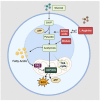
Basic metabolic regulation in ILC2s. ILC2s primarily rely on OXPHOS for energy production via FAO, both at rest or during activation. When circulating ILC2s are activated, arginine is metabolized into amino acids via the action of arginase 1 (Arg1), while BCAAs are metabolized into acetyl-CoA, contributing to the glycolytic output via OXPHOS.
Figure 3
Figure 3
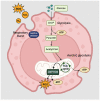
Basic metabolic regulation in eosinophils. Eosinophils have highly plastic metabolic capabilities depending on the environment and stimuli. It generates energy in the form of ATP through the process of glucose oxidation, aerobic glycolysis, or OXPHOS. OXPHOS occurs in the mitochondria, producing ROS and a high yield of ATP. Respiratory burst occurs during eosinophil activation, releasing ROS such as O2, which is then converted into H2O2.

References

    1. Qiu S., Cai Y., Yao H., Lin C., Xie Y., Tang S., Zhang A. Small molecule metabolites: Discovery of biomarkers and therapeutic targets. Signal Transduct. Target. Ther. 2023;8:132. - PMC - PubMed
    1. Baker S.A., Rutter J. Metabolites as signalling molecules. Nat. Rev. Mol. Cell Biol. 2023;24:355–374. doi: 10.1038/s41580-022-00572-w. - DOI - PubMed
    1. Li F., Xu W., Zhao S. Regulatory roles of metabolites in cell signaling networks. J. Genet. Genom. 2013;40:367–374. doi: 10.1016/j.jgg.2013.05.002. - DOI - PubMed
    1. Samitas K., Carter A., Kariyawasam H.H., Xanthou G. Upper and lower airway remodelling mechanisms in asthma, allergic rhinitis and chronic rhinosinusitis: The one airway concept revisited. Allergy. 2018;73:993–1002. doi: 10.1111/all.13373. - DOI - PubMed
    1. Qin Z., Chen Y., Wang Y., Xu Y., Liu T., Mu Q., Huang C. Immunometabolism in the pathogenesis of asthma. Immunology. 2024;171:1–17. doi: 10.1111/imm.13688. - DOI - PubMed

LinkOut - more resources