. 2024 Jul 5;25(13):7404.
doi: 10.3390/ijms25137404.

Piezo 1 and Piezo 2 in the Chemosensory Organs of Zebrafish (Danio rerio)

Affiliations

Piezo 1 and Piezo 2 in the Chemosensory Organs of Zebrafish (Danio rerio)

Marialuisa Aragona et al. Int J Mol Sci. .

Abstract

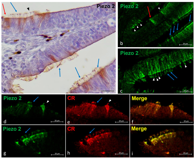
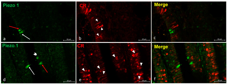
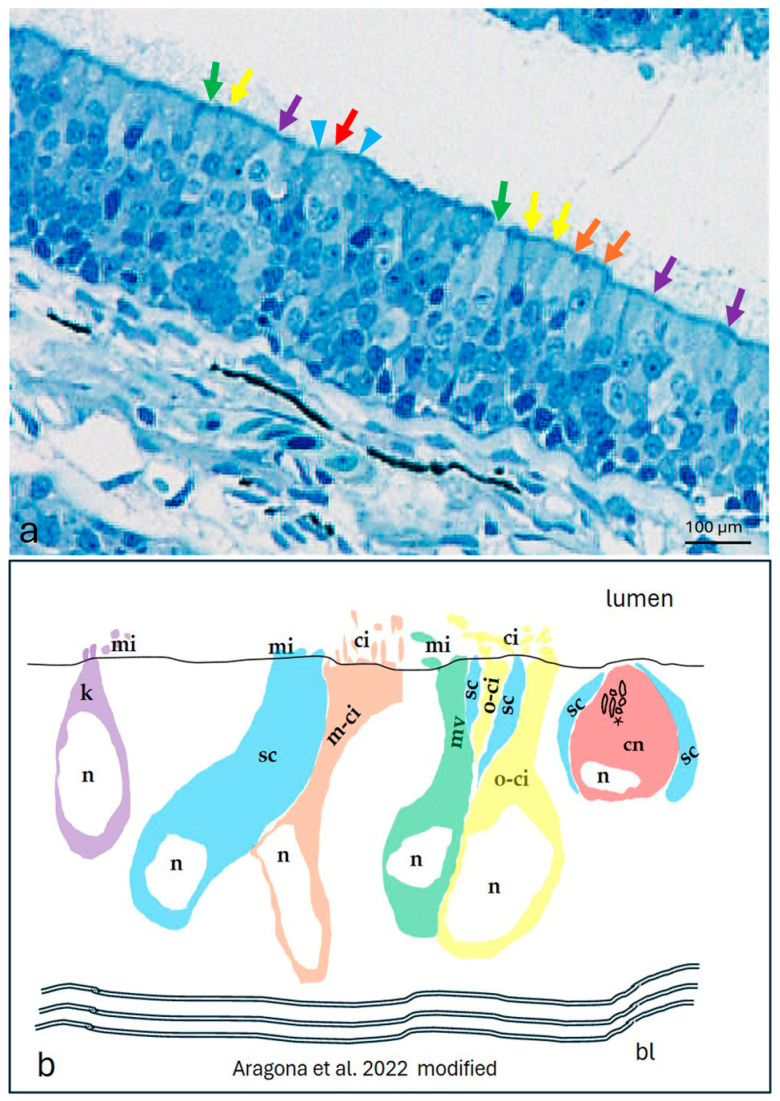
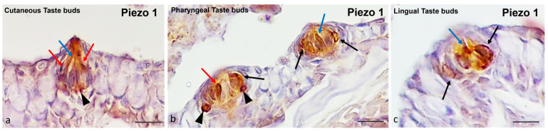
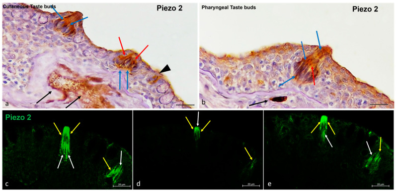
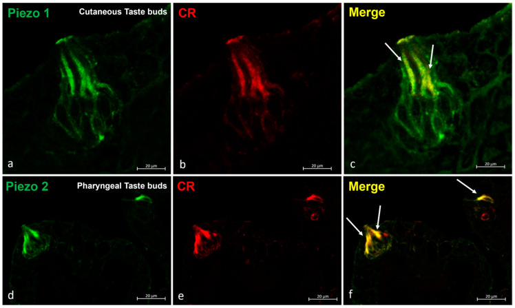
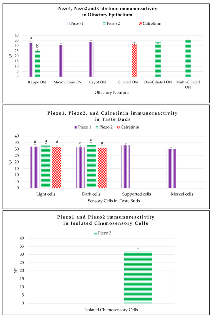
The ion channels Piezo 1 and Piezo 2 have been identified as membrane mechano-proteins. Studying mechanosensitive channels in chemosensory organs could help in understanding the mechanisms by which these channels operate, offering new therapeutic targets for various disorders. This study investigates the expression patterns of Piezo proteins in zebrafish chemosensory organs. For the first time, Piezo protein expression in adult zebrafish chemosensory organs is reported. In the olfactory epithelium, Piezo 1 immunolabels kappe neurons, microvillous cells, and crypt neurons, while Calretinin is expressed in ciliated sensory cells. The lack of overlap between Piezo 1 and Calretinin confirms Piezo 1's specificity for kappe neurons, microvillous cells, and crypt neurons. Piezo 2 shows intense immunoreactivity in kappe neurons, one-ciliated sensory cells, and multi-ciliated sensory cells, with overlapping Calretinin expression, indicating its olfactory neuron nature. In taste buds, Piezo 1 immunolabels Merkel-like cells at the bases of cutaneous and pharyngeal taste buds and the light and dark cells of cutaneous and oral taste buds. It also marks the dark cells of pharyngeal taste buds and support cells in oral taste buds. Piezo 2 is found in the light and dark cells of cutaneous and oral taste buds and isolated chemosensory cells. These findings provide new insights into the distribution of Piezo channels in zebrafish chemosensory organs, enhancing our understanding of their sensory processing and potential therapeutic applications.

Keywords: Piezo 1; Piezo 2; isolated chemosensory cells; olfactory epithelium; sensory organ; taste buds; translational medicine; zebrafish.


Conflict of interest statement

The authors declare no conflicts of interest.

Figures

Figure 4
Figure 4
Piezo 2 immunolocalizations in zebrafish olfactory lamellae, dorsal view. (a) The immunohistochemical detection (using a peroxidase method, Haematoxylin-stained) of Piezo 2 showing kappe neurons (red arrow), one-ciliated sensory cells (arrowhead), and multi-ciliated sensory cells (blue arrows). (b,c) The Piezo 2 immunolocalization showing kappe neurons (red arrow), one-ciliated sensory cells (arrowhead), and multi-ciliated sensory cells (blue arrows). (d,e,g,h) The immunohistochemical detection of Piezo 2 and Calretinin showing one-ciliated (arrowhead) and multi-ciliated (arrows) sensory cells. (f,i) A Piezo 2–Calretinin colocalization view. Magnification, 40×; scale bar, 20 µm.
Figure 1
Figure 1
Western blot analyses showing bands corresponding to the molecular weights of the zebrafish Piezo 1 (a) and Piezo 2 (b) proteins.
Figure 2
Figure 2
(a–f) Zebrafish olfactory lamellae, dorsal view. We conducted an immunohistochemical detection (using the immunofluorescences method) of Piezo 1 in a colocalization view with Calretinin as a specific marker for the neuronal subpopulation cells. (a,d) The kappe cells (red arrows) and microvillous sensory cells (white arrows) were immunopositive to Piezo 1. (d) Piezo 1 was localized in crypt neurons (gallon arrow). (b,e) The ciliated sensory cells were immunolabeled to Calretinin (arrowhead). (c,f) A colocalization view that shows no overlap in the labelling. Magnification, 40×; scale bar, 20 µm.
Figure 3
Figure 3
The olfactory epithelium of an adult zebrafish. (a) Photomicrographs of the semithin section showing the kappe cells (purple arrows), one-ciliated ON (yellow arrows), multi-ciliated ON (orange arrows), microvillous ON (green arrows), and crypt neurons (red arrow) surrounded by special supporting cells (blue arrowheads). Toluidine blue; magnification, 20×. (b) A zebrafish epithelium olfactory neuron’s graphic representation modified using the transmission electron microscopy micrograph from our previous study [10]. Abbreviations: k, kappe cells; sc, supporting cells; m-ci, multi-ciliated ON; n, nucleus; o-ci, one-ciliated ON, ci, cilium; mv, microvillous ON; mi, microvillus; cn, crypt neurons, with several cilia within the crypt (asterisk), special supporting cells (sc), and basal lamina (bl).
Figure 5
Figure 5
Zebrafish taste buds, transversal view. The immunohistochemical detection (using a peroxidase method) of Piezo 1. (a) In the cutaneous taste buds, the Merkel-like cells (arrowheads), light cells (blue arrows), and dark cells (red arrows) showed immunopositivity to Piezo 1. (b) In the pharyngeal taste buds, the Merkel-like cells (arrowheads), light cells (blue arrows), dark cells (red arrows), and supporting cells (black arrows) showed immunoreactivity to Piezo 1. (c) In the lingual taste buds, the light cells (blue arrow) and supporting cells (black arrows) were Piezo 1-immunolabelled. Magnification, 40×; scale bar, 20 µm.
Figure 6
Figure 6
Zebrafish taste buds, transversal view. (a,b) The immunohistochemical detection (using a peroxidase method, Haematoxylin-stained) of Piezo 2. The light cells (blue arrows), dark cells (red arrows), and nerve cells (black arrows) were Piezo 2-immunopositive. (a) Isolated chemosensory cells (black arrowhead) were immunoreactive to Piezo 2. (ce) The multiple focal planes of the Piezo 2 immunohistochemical detections (using a fluorescence method). The light cells (white arrows) and dark cells (yellow arrows) were immunoreactive to Piezo 2. Magnification, 40×; scale bar, 20 µm.
Figure 7
Figure 7
Zebrafish taste buds, transversal view. The immunohistochemical detection (using fluorescence methods) of Piezo 1 and Piezo 2. (ac) The double experiments with Piezo 1 and Calretinin showed an overlapping stain in the sensory cells (arrows) of the cutaneous taste buds. (df) The double experiments for Piezo 2 and Calretinin showed an overlapping stain in the sensory cells (arrows) of the pharyngeal taste buds. Magnification, 40×; scale bar, 20 µm.
Figure 8
Figure 8
Graphical representation of the immunoreactive cell counts: the kappe olfactory neurons (Kappe ON), microvillous olfactory neurons (Microvillous ON), crypt olfactory neurons (Crypt ON), one-ciliated olfactory neurons (One-ciliated ON), and multi-ciliated olfactory neurons (Multi-ciliated ON) in the olfactory epithelium labeled by Piezo 1, Piezo 2, and Calretinin; the light cells, dark cells, Merkel-like cells, and supporting cells of the taste buds immunolabeled by Piezo 1, Piezo 2, and Calretinin; and the isolated chemosensory cells marked by Piezo 2. The statistical analysis showed different expression patterns for the investigated proteins in the different cellular subpopulations. N°, mean of the cells immunopositive to Piezo 1, Piezo 2, and Calretinin. The lowercase letters indicate the statistical significance between the different cell subpopulations, with p < 0.05.

Similar articles

Cited by

References

    1. Hansen A., Eckart Z. The peripheral olfactory organ of the zebrafish, Danio rerio: An ultrastructural study. Chem. Senses. 1998;23:39–48. doi: 10.1093/chemse/23.1.39. - DOI - PubMed
    1. Hansen A., Reutter K., Zeiske E. Taste bud development in the zebrafish, Danio rerio. Dev. Dyn. 2002;223:483–496. doi: 10.1002/dvdy.10074. - DOI - PubMed
    1. Korsching S.I., Argo S., Campenhausen H., Friedrich R.W., Rummrich A., Weth F. Olfaction in zebrafish: What does a tiny teleost tell us? Semin. Cell Dev. Biol. 1997;8:181–187. doi: 10.1006/scdb.1996.0136. - DOI - PubMed
    1. Whitfield T.T. Zebrafish as a model for hearing and deafness. J. Neurobiol. 2002;53:157–171. doi: 10.1002/neu.10123. - DOI - PubMed
    1. Bhattarai P., Cosacak M.I., Mashkaryan V., Demir S., Popova S.D., Govindarajan N., Brandt K., Zhang Y., Chang W., Ampatzis K. Neuron-glia interaction through Serotonin-BDNF-NGFR axis enables regenerative neurogenesis in Alzheimer’s model of adult zebrafish brain. PLoS Biol. 2020;18:e3000585. doi: 10.1371/journal.pbio.3000585. - DOI - PMC - PubMed

LinkOut - more resources