

Clinical Applications and Future Directions of Smartphone Fundus Imaging
- PMID: 39001285
- PMCID: PMC11240943
- DOI: 10.3390/diagnostics14131395
Clinical Applications and Future Directions of Smartphone Fundus Imaging
Abstract
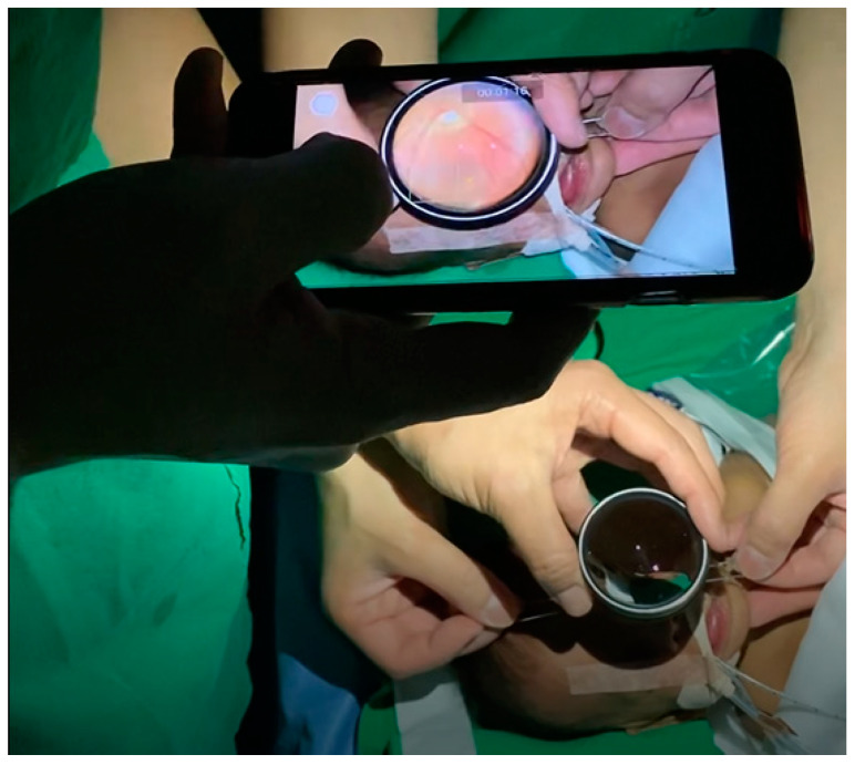
The advent of smartphone fundus imaging technology has marked a significant evolution in the field of ophthalmology, offering a novel approach to the diagnosis and management of retinopathy. This review provides an overview of smartphone fundus imaging, including clinical applications, advantages, limitations, clinical applications, and future directions. The traditional fundus imaging techniques are limited by their cost, portability, and accessibility, particularly in resource-limited settings. Smartphone fundus imaging emerges as a cost-effective, portable, and accessible alternative. This technology facilitates the early detection and monitoring of various retinal pathologies, including diabetic retinopathy, age-related macular degeneration, and retinal vascular disorders, thereby democratizing access to essential diagnostic services. Despite its advantages, smartphone fundus imaging faces challenges in image quality, standardization, regulatory considerations, and medicolegal issues. By addressing these limitations, this review highlights the areas for future research and development to fully harness the potential of smartphone fundus imaging in enhancing patient care and visual outcomes. The integration of this technology into telemedicine is also discussed, underscoring its role in facilitating remote patient care and collaborative care among physicians. Through this review, we aim to contribute to the understanding and advancement of smartphone fundus imaging as a valuable tool in ophthalmic practice, paving the way for its broader adoption and integration into medical diagnostics.
Keywords: clinical applications; fundus imaging; future directions; smartphone.
Conflict of interest statement
The authors declare no conflicts of interest.
Figures


References
-
- Panwar N., Huang P., Lee J., Keane P.A., Chuan T.S., Richhariya A., Teoh S., Lim T.H., Agrawal R. Fundus Photography in the 21st Century—A Review of Recent Technological Advances and Their Implications for Worldwide Healthcare. Telemed. J. e-Health. 2016;22:198–208. doi: 10.1089/tmj.2015.0068. - DOI - PMC - PubMed
-
- Patel T.P., Kim T.N., Yu G., Dedania V.S., Lieu P., Qian C.X., Besirli C.G., Demirci H., Margolis T., Fletcher D.A., et al. Smartphone-Based, Rapid, Wide-Field Fundus Photography for Diagnosis of Pediatric Retinal Diseases. Transl. Vis. Sci. Technol. 2019;8:29. doi: 10.1167/tvst.8.3.29. - DOI - PMC - PubMed
Publication types
LinkOut - more resources
Full Text Sources
