Interest of Integrated Whole-Body PET/MR Imaging in Gastroenteropancreatic Neuroendocrine Neoplasms: A Retro-Prospective Study
- PMID: 39001434
- PMCID: PMC11240462
- DOI: 10.3390/cancers16132372
Interest of Integrated Whole-Body PET/MR Imaging in Gastroenteropancreatic Neuroendocrine Neoplasms: A Retro-Prospective Study
Abstract
Introduction and aim: Simultaneous positron emission tomography/magnetic resonance imaging (PET-MRI) combines the high sensitivity of PET with the high specificity of MRI and is a tool for the assessment of gastroenteropancreatic neuroendocrine neoplasms (G-NENs). However, it remains poorly evaluated with no clear recommendations in current guidelines. Thus, we evaluated the prognostic impact of PET-MRI in G-NEN patients.
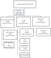
Methods: From June 2017 to December 2021, 71 G-NEN patients underwent whole-body PET-MRI for staging and/or follow-up purposes. A whole-body emission scan with 18F-6-fluoro-L-dihydroxyphenylalanine (18FDOPA, n = 30), 18F-fluoro-2-deoxy-D-glucose (18FDG, n = 21), or 68Ga-(DOTA(0)-Phe(1)-Tyr(3))-octreotide (68Ga-DOTATOC, n = 20) with the simultaneous acquisition of a T1-Dixon sequence and diffusion-weighed imaging (DWI), followed by a dedicated step of MRI sequences with a Gadolinium contrast was performed. The patients underwent PET-MRI every 6-12 months during the follow-up period until death. Over this period, 50 patients with two or more PET-MRI were evaluated.
Results: The mean age was 61 [extremes, 31-92] years. At the baseline, PET-MRI provided new information in 12 cases (17%) as compared to conventional imaging: there were more metastases in eight, an undescribed location (myocardia) in two, and an unknown primary location in two cases. G grading at the baseline influenced overall survival. During the follow-up (7-381 months, mean 194), clinical and therapy managements were influenced by PET-MRI in three (6%) patients due to new metastases findings when neither overall, nor disease-free survivals in these two subgroups (n = 12 vs. n = 59), were different.
Conclusion: Our study suggests that using PET/MRI with the appropriate radiotracer improves the diagnostic performance with no benefit on survival. Further studies are warranted to evaluate the cost-effectiveness of this procedure.
Keywords: G-NET; MRI; PET; PET-MRI; endocrine; gastrointestinal; pancreas.
Conflict of interest statement
The authors declare no conflicts of interest.
Figures


References
-
- Danti G., Flammia F., Matteuzzi B., Cozzi D., Berti V., Grazzini G., Pradella S., Recchia L., Brunese L., Miele V. Gastrointestinal Neuroendocrine Neoplasms (GI-NENs): Hot Topics in Morphological, Functional, and Prognostic Imaging. Radiol. Med. 2021;126:1497–1507. doi: 10.1007/s11547-021-01408-x. - DOI - PMC - PubMed
-
- Shah M.H., Goldner W.S., Benson A.B., Bergsland E., Blaszkowsky L.S., Brock P., Chan J., Das S., Dickson P.V., Fanta P., et al. Neuroendocrine and Adrenal Tumors, Version 2.2021, NCCN Clinical Practice Guidelines in Oncology. J. Natl. Compr. Cancer Netw. 2021;19:839–868. doi: 10.6004/jnccn.2021.0032. - DOI - PubMed
LinkOut - more resources
Full Text Sources

