. 2024 Jul 4;16(13):2456.
doi: 10.3390/cancers16132456.

Preliminary Experience in Ultra-High Frequency Ultrasound Assessment of Cutaneous Primary Lymphomas: An Innovative Classification

Affiliations

Preliminary Experience in Ultra-High Frequency Ultrasound Assessment of Cutaneous Primary Lymphomas: An Innovative Classification

Anna Russo et al. Cancers (Basel). .

Abstract

Background: Primary cutaneous lymphoma (PCL) is a rare form of extranodal non-Hodgkin's lymphoma characterized by malignant lymphocytes confined to the skin. Accurate diagnosis and staging are crucial for optimal management, yet radiological literature on imaging PCL remains limited. This study aims to delineate the imaging characteristics of PCLs using high and ultra-high frequency ultrasound (UHFUS) and proposes a classification system based on ultrasound findings.

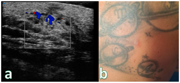
Methods: A cohort of 88 individuals with suspected PCL underwent high-resolution ultrasound (HRUS) and color Doppler examination of lesions. Lesions were categorized based on sonographic appearance, and subsequent histopathological assessment confirmed the diagnosis.

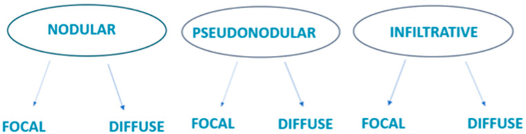
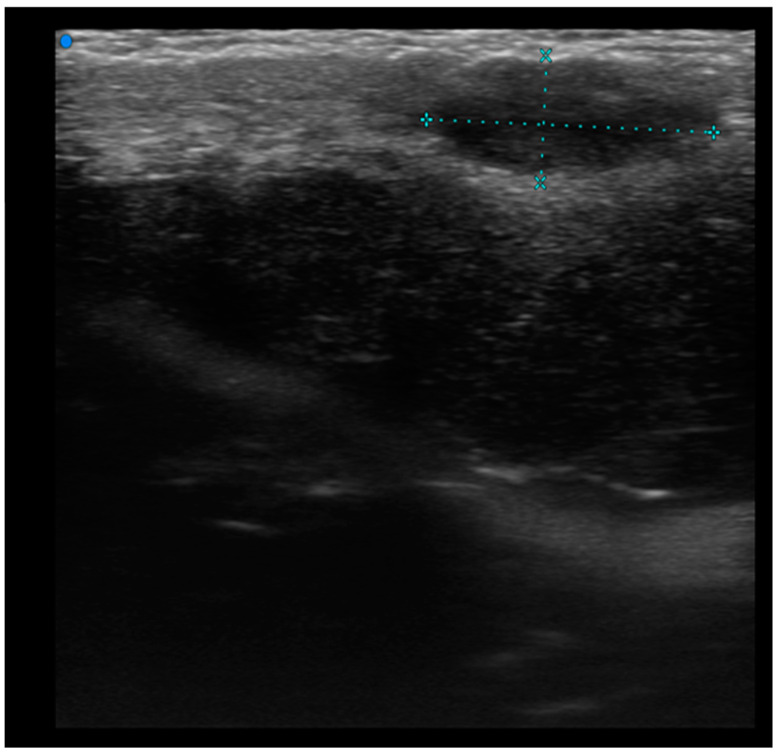
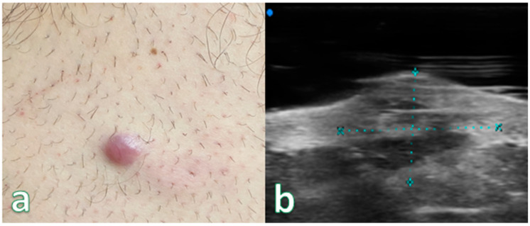
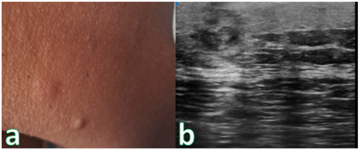
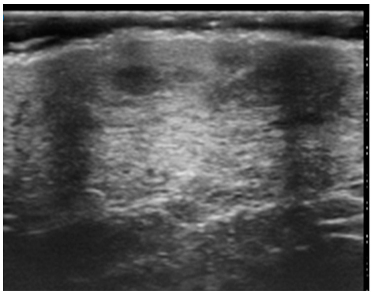
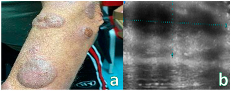
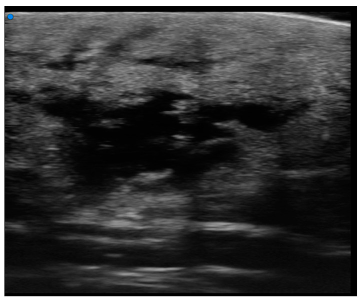
Results: Ultrasound imaging revealed distinct patterns for primary cutaneous T-cell lymphomas (PCTCL) and primary cutaneous B-cell lymphomas (PCBCL), with characteristic features such as hypoechoic nodules, pseudonodular lesions, and dermal infiltration. Histopathological analysis confirmed the ultrasound findings, supporting the proposed classification system.

Conclusions: Ultrasonography, particularly UHFUS, offers valuable insights into the imaging characteristics of primary cutaneous lymphomas, aiding the accurate diagnosis and assessment of treatment response. The proposed classification system based on ultrasound findings enhances the diagnostic approach to PCLs, and paves the way for improved patient care and management strategies.

Keywords: high-frequency ultrasound; oncologic ultrasonography; primary cutaneous lymphoma; ultra-high frequency ultrasound.


Conflict of interest statement

The authors declare no conflicts of interest.

Figures

Figure 1
Figure 1
In our experiment, we developed a new classification comprising six ultrasound patterns.
Figure 2
Figure 2
HFUS assessment of a round hypoechoic subepidermal nodular lesion showing no infiltration of deeper planes. Pathological assessment revealed it to be a B cell Primary Cutaneous Lymphoma.
Figure 3
Figure 3
(a) Reddish nodular skin lesion without signs of flogisis in the anterior region of the right leg. (b) Examination performed with a very high-frequency ultrasound probe at 50 MHz showing iso-hypoechoic lesion with irregular and hyperechoic margins infiltrating the epidermis and the subepidermal layer highlighted with dashed blue line. Final pathologic assessment revealed to be a B-Cell Lymphoma.
Figure 4
Figure 4
(a) Nodular skin lesion without signs of flogisis in the right deltoid region. (b) Exam performed with a very high-frequency ultrasound probe at 50 MHz: Iso-hypoechoic lesion with irregular and hyperechoic margins infiltrating the epidermis and the subepidermal layer. Pathologic Assessment revealed B-Cell Lymphoma.
Figure 5
Figure 5
Focal pseudonodular lesion (T-cell lymphoma). Examination performed with a very high-frequency ultrasound probe at 48 MHz in the region of the right scapula: Iso-hypoechoic lesions with regular margins and epidermal localization.
Figure 6
Figure 6
(a) Multiple pseudonodular lesion of the left forearm. (b) Examination performed with a very high-frequency ultrasound probe at 50 MHz in the anterior region of the left forearm showing iso-hypoechoic lesions with irregular margins located in the epidermal and subdermal layers highlighted by dashed blue lines. Images are low quality due to irregular surfaces of lesion.
Figure 7
Figure 7
Diffuse infiltrative lesion (recurrence of B-cell lymphoma) examination performed with a very high-frequency ultrasound probe at 50 MHz in the region of the right scapula: Iso-hypoechoic lesions with nodular and multilobulated morphology and irregular margins infiltrating the epidermal and subepidermal tissue.
Figure 8
Figure 8
(a) Examination performed with a very high-frequency ultrasound probe at 48 MHz, showing iso-hypoechoic lesions with nodular and multilobulated morphology and irregular margins infiltrating the epidermal and subepidermal tissue at a single point (focal). (b) Reddish, indistinct, slightly exofitic nodule in the lumbar region. Color Doppler analysis showing no significative increase in the signals. Pathological assessment showed B-Cell Lymphoma (recurrence).

Similar articles

Cited by

References

    1. Lebas E., Collins P., Somja J., Nikkels A.F. A Comprehensive Update of the Atypical, Rare and Mimicking Presentations of Mycosis Fungoides. Dermatol. Ther. 2021;11:1931–1951. doi: 10.1007/s13555-021-00625-6. - DOI - PMC - PubMed
    1. Sonego B., Ibatici A., Rivoli G., Angelucci E., Sola S., Massone C. Histopathological Markers for Target Therapies in Primary Cutaneous Lymphomas. Cells. 2023;12:2656. doi: 10.3390/cells12222656. - DOI - PMC - PubMed
    1. Roccuzzo G., Giordano S., Fava P., Pileri A., Guglielmo A., Tonella L., Sanlorenzo M., Ribero S., Fierro M.T., Quaglino P. Immune Check Point Inhibitors in Primary Cutaneous T-Cell Lymphomas: Biologic Rationale, Clinical Results and Future Perspectives. Front. Oncol. 2021;11:733770. doi: 10.3389/fonc.2021.733770. - DOI - PMC - PubMed
    1. Piccolo V., Mascolo M., Russo T., Staibano S., Argenziano G. Dermoscopy of primary cutaneous B-cell lymphoma (PCBCL) J. Am. Acad. Dermatol. 2016;75:e137–e139. doi: 10.1016/j.jaad.2016.02.1217. - DOI - PubMed
    1. Olsen E.A., Whittaker S., Willemze R., Pinter-Brown L., Foss F.M., Geskin L.J., Schwartz L.H., Horwitz S.M., Guitart J., Zic J., et al. Primary cutaneous lymphoma: Recommendations for clinical trial design and staging update from the ISCL, USCLC, and EORTC. Blood. 2022;140:419–437. doi: 10.1182/blood.2021012057. - DOI - PMC - PubMed

LinkOut - more resources