

Differential Diagnosis of Pigmented Lesions in the Oral Mucosa: A Clinical Based Overview and Narrative Review
- PMID: 39001549
- PMCID: PMC11240708
- DOI: 10.3390/cancers16132487
Differential Diagnosis of Pigmented Lesions in the Oral Mucosa: A Clinical Based Overview and Narrative Review
Abstract
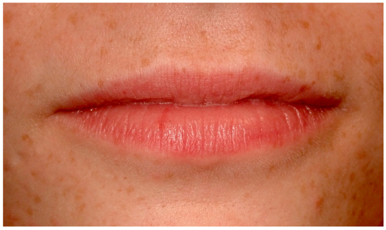
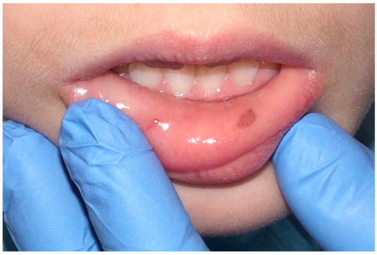
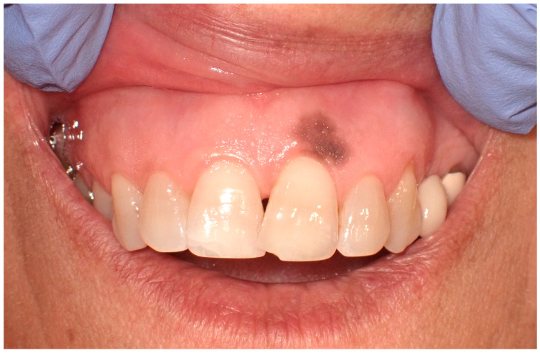
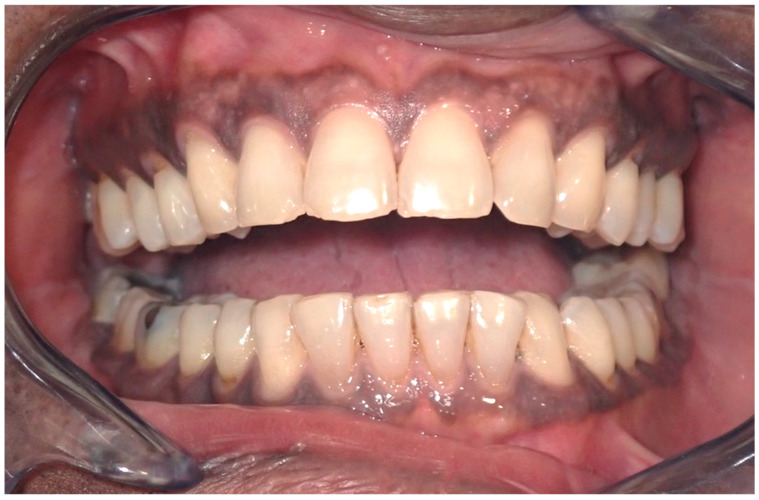
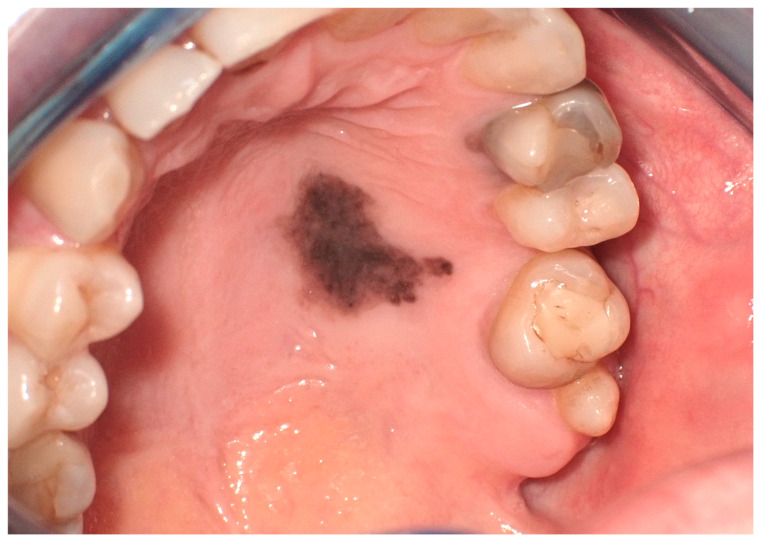
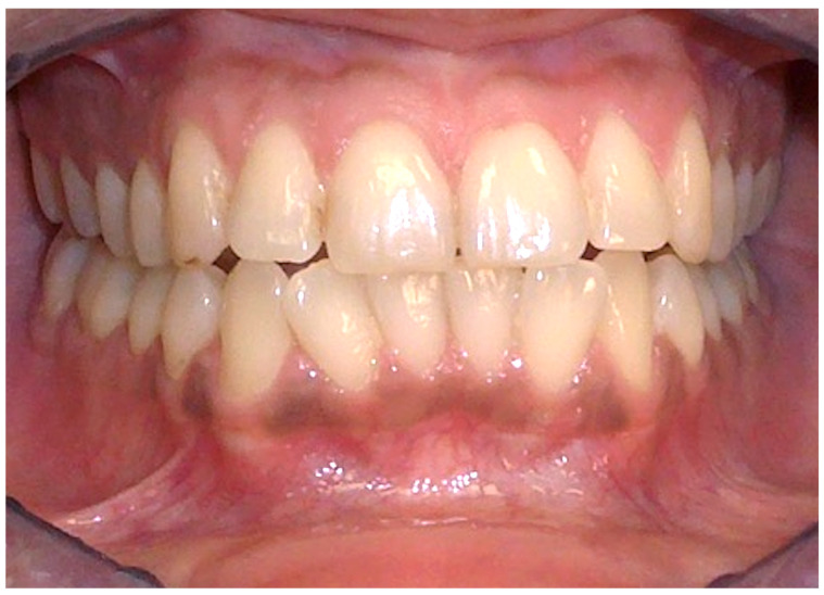
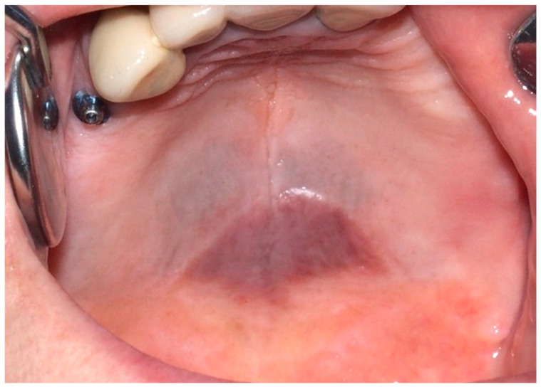
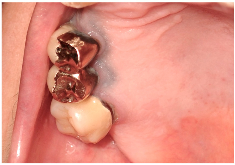
This paper examines the clinical differentiation of pigmented lesions in the oral mucosa, which poses significant diagnostic challenges across dental and medical disciplines due to their spectrum from benign to potentially malignant conditions. Through a literature review and analysis of clinical cases, this study clarifies current diagnostic methodologies, with an emphasis on differential diagnosis, to provide a practical guide for clinicians. The classification of pigmented lesions, such as endogenous, focal melanocytic, and multifocal pigmentation, based on clinical and histological features, highlights the necessity for a structured and informed approach. A retrospective examination of cases from our oral medicine and pathology clinic, coupled with analysis of photographic and histological records, aids in classifying these lesions. This fosters a better understanding and promotes informed discussions among clinicians, ultimately aiming to enhance early and precise diagnosis, thus improving patient management and outcomes.
Keywords: malignant melanoma; melanin; mouth mucosa; oral health; oral medicine; oral pathology; oral pigmentation; pigmented lesions.
Conflict of interest statement
The authors declare no conflicts of interest.
Figures
Similar articles
-
Diagnosis of oral pigmentations and malignant transformations.Singapore Dent J. 2014 Dec;35C:39-46. doi: 10.1016/j.sdj.2014.03.001. Singapore Dent J. 2014. PMID: 25496584 Review.
-
Pigmented Lesions of the Oral Mucosa: Clinical Presentation, Histology, and Recommendations for Management.Am J Clin Dermatol. 2025 May 14. doi: 10.1007/s40257-025-00950-y. Online ahead of print. Am J Clin Dermatol. 2025. PMID: 40369390 Review.
-
[Oral medicine 10. Pigmented lesions of the oral mucosa].Ned Tijdschr Tandheelkd. 2013 Oct;120(10):555-61. Ned Tijdschr Tandheelkd. 2013. PMID: 25026743 Review. Dutch.
-
[A colorimetric study of pigmented lesions on oral mucosa].Kokubyo Gakkai Zasshi. 2010 Jun;77(2):149-55. Kokubyo Gakkai Zasshi. 2010. PMID: 20662308 Japanese.
-
Oral pigmented lesions: a retrospective analysis from Brazil.Med Oral Patol Oral Cir Bucal. 2021 May 1;26(3):e284-e291. doi: 10.4317/medoral.24168. Med Oral Patol Oral Cir Bucal. 2021. PMID: 32856618 Free PMC article.
Cited by
-
Prevalence and associated factors of oral pigmented lesions among Yemeni dental patients: a large cross-sectional study.BMC Oral Health. 2025 Mar 15;25(1):391. doi: 10.1186/s12903-025-05760-6. BMC Oral Health. 2025. PMID: 40089755 Free PMC article.
References
-
- Carson Dewitt R. The Gale Encyclopedia of Dermatology. 1st ed. Gale; Farmington Hills, MI, USA: 2017. pp. 244–246.
Publication types
LinkOut - more resources
Full Text Sources
