. 2024 Jul 8;16(13):2490.
doi: 10.3390/cancers16132490.

Transcriptomic Analysis Reveals Early Alterations Associated with Intrinsic Resistance to Targeted Therapy in Lung Adenocarcinoma Cell Lines

Affiliations

Transcriptomic Analysis Reveals Early Alterations Associated with Intrinsic Resistance to Targeted Therapy in Lung Adenocarcinoma Cell Lines

Mario Perez-Medina et al. Cancers (Basel). .

Abstract

Lung adenocarcinoma is the most prevalent form of lung cancer, and drug resistance poses a significant obstacle in its treatment. This study aimed to investigate the overexpression of long non-coding RNAs (lncRNAs) as a mechanism that promotes intrinsic resistance in tumor cells from the onset of treatment. Drug-tolerant persister (DTP) cells are a subset of cancer cells that survive and proliferate after exposure to therapeutic drugs, making them an essential object of study in cancer treatment. The molecular mechanisms underlying DTP cell survival are not fully understood; however, long non-coding RNAs (lncRNAs) have been proposed to play a crucial role. DTP cells from lung adenocarcinoma cell lines were obtained after single exposure to tyrosine kinase inhibitors (TKIs; erlotinib or osimertinib). After establishing DTP cells, RNA sequencing was performed to investigate the differential expression of the lncRNAs. Some lncRNAs and one mRNA were overexpressed in DTP cells. The clinical relevance of lncRNAs was evaluated in a cohort of patients with lung adenocarcinoma from The Cancer Genome Atlas (TCGA). RT-qPCR validated the overexpression of lncRNAs and mRNA in the residual DTP cells and LUAD biopsies. Knockdown of these lncRNAs increases the sensitivity of DTP cells to therapeutic drugs. This study provides an opportunity to investigate the involvement of lncRNAs in the genetic and epigenetic mechanisms that underlie intrinsic resistance. The identified lncRNAs and CD74 mRNA may serve as potential prognostic markers or therapeutic targets to improve the overall survival (OS) of patients with lung cancer.

Keywords: CD74; drug-tolerant persister (DTP) cells; erlotinib; intrinsic resistance; lung adenocarcinoma; osimertinib; senescent cells; tyrosine kinase inhibitors (TKIs).


Conflict of interest statement

The authors declare no conflicts of interest.

Figures

Figure 1
Figure 1
Morphological changes and proportion of cells in the cell-cycle phases in EGFR-mutated lung adenocarcinoma cell lines after a single erlotinib exposure. Cellular density changes induced by 200 nM erlotinib (A,D,G). Magnification 20×. Histograms showing the cell-cycle distribution profile and graphs of the percentage of cells in the cell-cycle phases induced by erlotinib in the HCC827 (B,C), HCC4006 (E,F), and H1975 (H,I) cell lines. Data are shown as the mean ± SD. **** p < 0.0001.
Figure 2
Figure 2
Effect of osimertinib exposure on EGFR-mutated lung adenocarcinoma cell lines. Morphological changes induced by single 200 nM osimertinib exposure (A,D,G). Magnification 20×. Histograms show the profile of cell distribution and graphs of the percentage of cells in the cell-cycle phases and the sub-G0 peak induced by osimertinib in the HCC827 (B,C), HCC4006 (E,F), and H1975 (H,I) cell lines. Data are shown as mean ± SD. **** p < 0.0001. (J). After a single 48 h exposure to osimertinib, residual cells from the H1975 cell line were cultured in fresh complete media for five days, and the expression of beta-galactosidase was detected. Micrographs show β-gal-positive cells. Magnification 40×.
Figure 3
Figure 3
Differentially expressed genes (DEGs) in DTP cells. Volcano plots showing DEGs in DTPs relative to untreated cells. Gray shading indicates overexpressed transcripts (A,B). Venn diagrams of the overexpressed genes shared among DTP cells (C,D). Functional annotation of the overexpressed lncRNAs was performed using the Gene Ontology (GO) biological process database (E,F).
Figure 4
Figure 4
Clinical relevance of lncRNAs and CD74 associated with intrinsic resistance. K–M survival analysis of the LUAD patient cohort from The Cancer Genome Atlas. High expression is shown in red, and low expression is shown in blue. Log-rank p values are indicated.
Figure 5
Figure 5
Gene expression of clinically relevant lncRNAs detected in DTP cells. Expression of the lncRNAs detected in the erlotinib- and osimertinib-induced DTP cells. Fold changes relative to the control are shown (A,B). DsiRNA-mediated knockdown reduced the expression of lncRNAs AGAP2-AS1 in the HCC827 cell line and LINC01133 and CD74 in the H1975 cell line (red). Mock (light blue) and DsiRNA against HPRT (green) were used as controls (C). Knockdown of the DTP-associated lncRNAs reduced cell viability after a single exposure to TKIs (D). Data are shown as mean ± SD of three independent experiments. **** p-value < 0.05.
Figure 6
Figure 6
Expression of clinically relevant lncRNAs and CD74 in tumor biopsies. Expression of DTP-associated genes in four FFPE lung adenocarcinoma biopsies detected using RT–qPCR. Gene expression was normalized to GAPDH and compared to that of Patient 1.
Figure 7
Figure 7
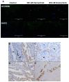
CD74 expression in lung adenocarcinoma. Immunofluorescence analysis of CD74 in osimertinib-induced DTP cells. CD74 mainly showed cytoplasmic staining, which increased in osimertinib-induced DTP cells. CD74 (green) and nuclear staining (blue). Magnification 20× (A). Lung adenocarcinoma biopsies. Comparison of CD74 staining between normal ciliated epithelial cells (star) and malignant cells (B). CD74-negative (arrow) and CD74-positive tumor cells within the same niche (C). Tumor cells expressing varying levels of CD74 (D). High CD74 staining was observed in tumor cells with invasive morphology (E). Magnification 20×.

Similar articles

Cited by

References

    1. Sung H., Ferlay J., Siegel R.L., Laversanne M., Soerjomataram I., Jemal A., Bray F. Global Cancer Statistics 2020: GLOBOCAN Estimates of Incidence and Mortality Worldwide for 36 Cancers in 185 Countries. CA. Cancer J. Clin. 2021;71:209–249. doi: 10.3322/caac.21660. - DOI - PubMed
    1. Tfayli A., Mohty R. EGFR Tyrosine Kinase Inhibitors in Non-Small Cell Lung Cancer: Treatment Paradigm, Current Evidence, and Challenges. Tumori J. 2020;107:376–384. doi: 10.1177/0300891620968138. - DOI - PubMed
    1. Jamal-Hanjani M., Wilson G.A., McGranahan N., Birkbak N.J., Watkins T.B.K., Veeriah S., Shafi S., Johnson D.H., Mitter R., Rosenthal R., et al. Tracking the Evolution of Non–Small-Cell Lung Cancer. N. Engl. J. Med. 2017;376:2109–2121. doi: 10.1056/NEJMoa1616288. - DOI - PubMed
    1. Nie N., Li J., Zhang J., Dai J., Liu Z., Ding Z., Wang Y., Zhu M., Hu C., Han R., et al. First-Line Osimertinib in Patients with EGFR-Mutated Non-Small Cell Lung Cancer: Effectiveness, Resistance Mechanisms, and Prognosis of Different Subsequent Treatments. Clin. Med. Insights Oncol. 2022;16:117955492211347. doi: 10.1177/11795549221134735. - DOI - PMC - PubMed
    1. Gharwan H., Groninger H. Kinase Inhibitors and Monoclonal Antibodies in Oncology: Clinical Implications. Nat. Rev. Clin. Oncol. 2016;13:209–227. doi: 10.1038/nrclinonc.2015.213. - DOI - PubMed

LinkOut - more resources