Myocardial Characteristics, Cardiac Structure, and Cardiac Function in Systemic Light-Chain Amyloidosis
- PMID: 39001736
- PMCID: PMC12009672
- DOI: 10.1016/j.jcmg.2024.05.004
Myocardial Characteristics, Cardiac Structure, and Cardiac Function in Systemic Light-Chain Amyloidosis
Abstract
Background: In systemic light-chain (AL) amyloidosis, cardiac involvement portends poor outcomes.
Objectives: The authors' objectives were to detect early myocardial alterations, to analyze longitudinal changes with therapy, and to predict major adverse cardiac events (MACE) in participants with AL amyloidosis using cardiac magnetic resonance imaging (MRI).
Methods: Recently diagnosed participants were prospectively enrolled. AL amyloidosis with and without cardiomyopathy (AL-CMP, AL-non-CMP) were defined based on abnormal cardiac biomarkers and wall thickness. MRI was performed at baseline, 6 months in all participants, and 12 months in participants with AL-CMP. MACE were defined as all-cause death, heart failure hospitalization, and cardiac transplantation. Mayo stage was based on troponin T, N-terminal pro-B-type natriuretic peptide, and difference in free light chains.
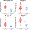
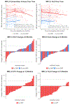
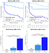
Results: This study included 80 participants (median age 62 years, 58% men). Extracellular volume (ECV) was abnormal (>32%) in all participants with AL-CMP and in 47% of those with AL-non-CMP. ECV tended to increase at 6 months (median +2%; AL-CMP P = 0.120; AL-non-CMP P = 0.018) and returned to baseline values at 12 months in participants with AL-CMP. Global longitudinal strain (GLS) improved at 6 months (median -0.6%; P = 0.048) and 12 months (median -1.2%; P < 0.001) in participants with AL-CMP. ECV and GLS were strongly associated with MACE (P < 0.001) and improved the prognostic value when added to Mayo stage (P ≤ 0.002). No participant with ECV ≤32% had MACE, while 74% of those with ECV >48% had MACE.
Conclusions: In patients with systemic AL amyloidosis, ECV detects subclinical myocardial alterations. With therapy, ECV tends to increase at 6 months and returns to values unchanged from baseline at 12 months, whereas GLS improves at 6 and 12 months in participants with AL-CMP. ECV and GLS offer additional prognostic performance over Mayo stage. (Molecular Imaging of Primary Amyloid Cardiomyopathy [MICA]; NCT02641145).
Keywords: T1 mapping; cardiac magnetic resonance imaging; extracellular volume; global longitudinal strain; light-chain (AL) amyloidosis; myocardial characterization.
Copyright © 2024 American College of Cardiology Foundation. Published by Elsevier Inc. All rights reserved.
Conflict of interest statement
Funding Support and Author Disclosures This work was supported by National Institutes of Health. Dr Dorbala was supported by grants R01 HL 130563, K24 HL 157648, AHA16 CSA 2888 0004, and AHA19SRG34950011. Dr Falk was supported by grant R01 HL 130563. Dr Liao was supported by grants AHA16 CSA 2888 0004 and AHA19SRG34950011. Dr Ruberg was supported by grants R01 HL 130563 and R01 HL 093148. Dr Cuddy was supported by grants NIH 1K23HL166686-01 and AHA 23CDA857664NIH. Dr Bianchi was partially supported by a grant K08 CA245100. Dr Clerc has received a research fellowship from the International Society of Amyloidosis and Pfizer. Dr Cuddy has received an investigator-initiated research grant from Pfizer, as well as consulting fees from BridgeBio, Ionis, Astra Zeneca and Novo Nordisk. Dr Bianchi has received consulting fees from Prothena. Dr Ruberg has received consulting fees from AstraZeneca and Attralus; and has received research support from Pfizer, Alnylam, Anumana, and Ionis/Akcea. Dr DiCarli has received a research grant from Spectrum Dynamics and Gilead; and has received consulting fees from Sanofi and General Electric. Dr Kwong has received grant funding from Alynlam Pharmaceuticals. Dr Falk has received consulting fees from Ionis Pharmaceuticals, Alnylam Pharmaceuticals, and Caelum Biosciences; and has received research funding from GlaxoSmithKline and Akcea. Dr Dorbala has received consulting fees from Pfizer, GE Healthcare, and Novo Nordisk; and has received investigator-initiated grants from Pfizer, GE Healthcare, Attralus, Siemens, and Phillips. All other authors have reported that they have no relationships relevant to the contents of this paper to disclose.
Figures


References
-
- Falk RH, Alexander KM, Liao R, Dorbala S. AL (Light-Chain) Cardiac Amyloidosis: A Review of Diagnosis and Therapy. J Am Coll Cardiol. 2016;68:1323–1341. - PubMed
-
- Dorbala S, Ando Y, Bokhari S, et al. ASNC/AHA/ASE/EANM/HFSA/ISA/SCMR/SNMMI expert consensus recommendations for multimodality imaging in cardiac amyloidosis: Part 1 of 2-evidence base and standardized methods of imaging. J Nucl Cardiol. 2019;26:2065–2123. - PubMed
Publication types
MeSH terms
Substances
Associated data
Grants and funding
LinkOut - more resources
Full Text Sources
Medical
Miscellaneous

