. 2024 Oct 14;120(12):1456-1471.
doi: 10.1093/cvr/cvae144.

Genetic deletion or pharmacologic inhibition of histone deacetylase 6 protects the heart against ischaemia/reperfusion injury by limiting tumour necrosis factor alpha-induced mitochondrial injury in experimental diabetes

Affiliations

Genetic deletion or pharmacologic inhibition of histone deacetylase 6 protects the heart against ischaemia/reperfusion injury by limiting tumour necrosis factor alpha-induced mitochondrial injury in experimental diabetes

Shelley L Baumgardt et al. Cardiovasc Res. .

Abstract

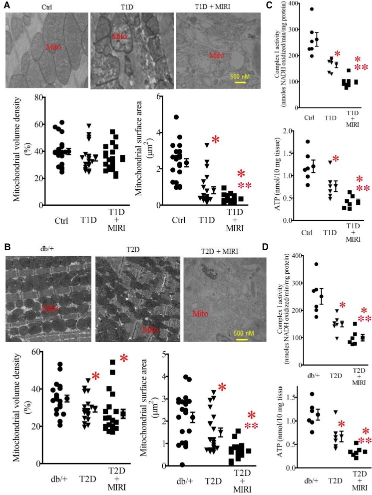
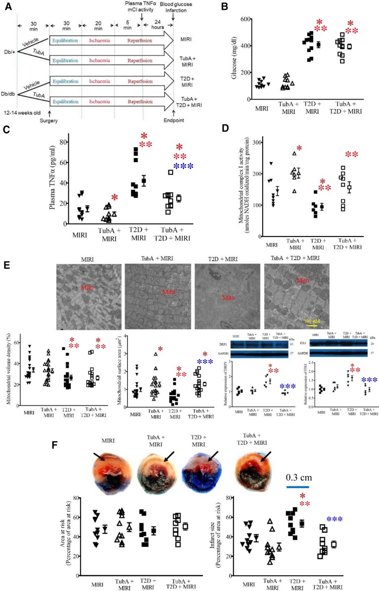
Aims: The histone deacetylase 6 (HDAC6) inhibitor, tubastatin A (TubA), reduces myocardial ischaemia/reperfusion injury (MIRI) in type 1 diabetic rats. It remains unclear whether HDAC6 regulates MIRI in type 2 diabetic animals. Diabetes augments the activity of HDAC6 and the generation of tumour necrosis factor alpha (TNF-α) and impairs mitochondrial complex I (mCI). Here, we examined how HDAC6 regulates TNF-α production, mCI activity, mitochondria, and cardiac function in type 1 and type 2 diabetic mice undergoing MIRI.

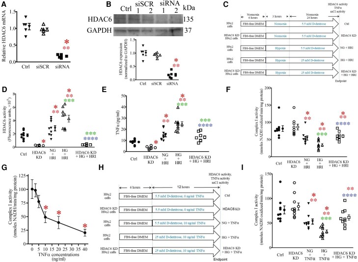
Methods and results: HDAC6 knockout, streptozotocin-induced type 1 diabetic, and obese type 2 diabetic db/db mice underwent MIRI in vivo or ex vivo in a Langendorff-perfused system. We found that MIRI and diabetes additively augmented myocardial HDAC6 activity and generation of TNF-α, along with cardiac mitochondrial fission, low bioactivity of mCI, and low production of adenosine triphosphate. Importantly, genetic disruption of HDAC6 or TubA decreased TNF-α levels, mitochondrial fission, and myocardial mitochondrial nicotinamide adenine dinucleotide levels in ischaemic/reperfused diabetic mice, concomitant with augmented mCI activity, decreased infarct size, and improved cardiac function. Moreover, HDAC6 knockout or TubA treatment decreased left ventricular dilation and improved cardiac systolic function 28 days after MIRI. H9c2 cardiomyocytes with and without HDAC6 knockdown were subjected to hypoxia/reoxygenation injury in the presence of high glucose. Hypoxia/reoxygenation augmented HDAC6 activity and TNF-α levels and decreased mCI activity. These negative effects were blocked by HDAC6 knockdown.

Conclusion: HDAC6 is an essential negative regulator of MIRI in diabetes. Genetic deletion or pharmacologic inhibition of HDAC6 protects the heart from MIRI by limiting TNF-α-induced mitochondrial injury in experimental diabetes.

Keywords: Histone deacetylase 6; Ischaemia/reperfusion; Mitochondria; Tumour necrosis factor alpha; Type 1 diabetes; Type 2 diabetes.


Conflict of interest statement

Conflict of interest: none declared.

Figures

Graphical Abstract
Graphical Abstract
Figure 1
Figure 1
Cardiac HDAC6 activity was enhanced in T1D mice undergoing sham surgery and MIRI. (A) Experimental procedures for STZ-induced T1D mice. (B) Fasting blood glucose in T1D and Ctrl mice 24 h after ischaemia or sham surgery. (C) Top: representative heart sections stained with 2,3,5-TTC and phthalocyanine blue showing area at risk and infarct size . The area at risk was defined as regions not stained with phthalocyanine blue dye, and the infarct area was defined as regions not stained with 2,3,5-TTC. Bottom: infarct size expressed as a percentage of area at risk in T1D and Ctrl mice. (D) Cardiac HDAC6 activity was enhanced in T1D and T1D + MIRI mice. Data are presented as means ± SEM. Kruskal–Wallis test followed by Dunn's test was used to analyse multiple group comparisons. *P < 0.05 vs. Ctrl and **P < 0.05 vs. T1D (n = 9–10 mice/group). (E) Experimental procedures for obese T2D db/db mice and controls (db/+). (F) Fasting blood glucose in db/db mice and Ctrl 24 h after ischaemia or sham surgery. (G) Top: heart sections stained with TTC and phthalocyanine blue showing area at risk and infarct size. Bottom: infarct size expressed as a percentage of area at risk in T2D and Ctrl mice. (H) Cardiac HDAC6 activity was increased in T2D and T2D + MIRI db/+ mice. Kruskal–Wallis test followed by Dunn's test was used to analyse multiple group comparisons. *P < 0.05 vs. db/+ and **P < 0.05 vs. T2D (n = 9–10 mice/group). Scale bar: 0.3 cm.
Figure 2
Figure 2
Mitochondrial morphology, mCI activity, and ATP contents in T1D and T2D mice undergoing sham surgery and MIRI. (A) Top: representative electron microscope micrographs showing the changes in mitochondria of T1D mice and controls. Bottom: mitochondrial volume density (n = 20–21 sections from 3 mice/group) and mitochondrial surface area (n = 200–210 mitochondria from 3 mice/group). Ctrl, non-diabetic C57BL/6 mice were subjected to sham surgery; T1D, type 1 diabetic C57BL/6 mice underwent sham surgery; T1D + MIRI, diabetic C57BL/6 mice underwent 20 min of ischaemia followed by reperfusion for 24 h. (B) Top: representative electron microscope micrographs showing the changes in mitochondria of T2D mice and controls. Bottom: mitochondrial volume density (n = 20–21 sections from 3 mice/group) and mitochondrial surface area (n = 200–210 mitochondria from 3 mice /group). Db/+, non-diabetic db/+ mice were subjected to sham surgery; T2D, type 2 diabetic db/db mice underwent sham surgery; T2D + MIRI; db/db mice underwent 20 min of ischaemia followed by reperfusion for 24 h. (C) T1D and MIRI jointly decreased mCI activity and ATP contents. Myocardial mCI activity 5 min after post-ischaemic reperfusion (n = 6–8 hearts/group) and myocardial ATP contents (n = 6 hearts/group) in T1D mice and Ctrl mice. Data are presented as means ± SEM. Kruskal–Wallis test followed by Dunn's test was used to analyse multiple group comparisons. *P < 0.05 vs. Ctrl, **P < 0.05 vs. T1D groups. (D) T2D and MIRI jointly decreased mCI activity and ATP contents. Myocardial mCI activity 5 min after reperfusion (n = 6–8 hearts/group) and myocardial ATP contents (n = 6 hearts/group) in T2D mice and Ctrl mice. Scale bar: 500 nm.
Figure 3
Figure 3
HDAC6 KO decreased plasma TNF-α and infarct size and augmented mCI activity during reperfusion in type 1 diabetic mice. (A) Experimental procedures. (B) Fasting blood glucose in HDAC6−/− and C57BL/6 mice. (C) HDAC6 KO decreased plasma TNF-α levels in T1D mice undergoing MIRI. (D) HDAC6 KO increased mCI activity in T1D mice undergoing MIRI (n = 9–11 hearts/group). (E) HDAC6 KO increased mitochondrial surface area and decreased the expression of cardiac DRP1 and FIS1 in T1D mice undergoing MIRI. Top: representative electron microscope micrographs of mitochondria. Bottom: mitochondrial volume density (n = 20–21 sections from 3 mice/group), mitochondria surface area (n = 200–210 mitochondria from 3 mice/group), and the expression of cardiac DRP1 and FIS1 (n = 5 hearts/group). Scale bar: 500 nm. (F) HDAC6 KO decreased myocardial infarct size in T1D mice. Top: representative heart images showing area at risk (non-black area) and infarct size. Arrows point to infarct area (white). Scale bar: 0.3 cm. Bottom: area at risk expressed as a percentage of the left ventricle and infarct size expressed as a percentage of area at risk (n = 8–10 mice/group). Data are presented as means ± SEM. Kruskal–Wallis test followed by Dunn's test was used to analyse multiple group comparisons. *P < 0.05 vs. MIRI groups, **P < 0.05 vs. HDAC6−/− + MIRI groups, ***P < 0.05 vs. T1D + MIRI groups.
Figure 4
Figure 4
Inhibition of HDAC6 decreased plasma TNF-α and infarct size and augmented mCI activity during reperfusion in T2D mice. (A) Experimental procedures. (B) Fasting blood glucose in db/db mice and db/+ control mice. (C) TubA decreased plasma TNF-α levels in T2D mice undergoing MIRI. (D) TubA enhanced myocardial mCI activity in T2D mice undergoing MIRI. (E) TubA increases mitochondrial size and decreased the expression of cardiac DRP1 and FIS1 in T2D mice undergoing MIRI. Top: representative electron microscope micrographs of mitochondria. Bottom: mitochondrial volume density (n = 20–21 sections from 3 mice/group), mitochondria surface area (n = 200–210 mitochondria from 3 mice/group), and the expression of cardiac DRP1 and FIS 1 (n = 5 hearts/group). Scale bar: 500 nm. (F) TubA decreased myocardial infarct size in T2D mice. Top: representative heart images showing area at risk and infarct size. Arrows point to infarct area. Scale bar: 0.3 cm. Bottom: area at risk expressed as a percentage of the left ventricle and infarct size expressed as a percentage of area at risk (n = 8–10 mice/group). Data are presented as means ± SEM. Kruskal–Wallis test followed by Dunn's test was used to analyse multiple group comparisons. *P < 0.05 vs. MIRI groups, **P < 0.05 vs. TubA + MIRI groups, ***P < 0.05 vs. T2D + MIRI groups.
Figure 5
Figure 5
HDAC6 KO reduced T1D-elicited increases in mitochondrial NAD(H) levels and improved cardiac function during reperfusion in Langendorff-perfused hearts. (A) Schematic presentation of experimental procedure of ex vivo experiments for measurements of NAD(H) and cardiac function of HDAC6−/− and C57BL/6 mice undergoing ischaemia/reperfusion injury (MIRI; n = 8–9 mice/group). (B) The dynastic changes in NAD(H) fluorescence of Langendorff-perfused mouse hearts at baseline, ischaemia, and reperfusion. (C) LV end-diastolic function. (D) LVDP. (E) +dP/dt (maximum rate of increase of LVDP). (F) −dP/dt (maximum rate of decrease of LVDP). Data are presented as means ± SEM. Kruskal–Wallis test followed by Dunn's test was used to analyse multiple group comparisons. *P < 0.05 vs. MIRI groups, **P < 0.05 vs. HDAC6−/− + MIRI groups, ***P < 0.05 vs. T1D + MIRI.
Figure 6
Figure 6
HDAC6 KD blocked TNF-α–induced inhibition of mCI activity in cardiomyocytes. (A) Quantitative reverse transcriptase-polymerase chain reaction analysis of HDAC6 mRNA levels in H9c2 cardiomyocytes. (B) Western blot analysis of HDAC6 protein expression in H9c2 cardiomyocytes. Top: representative western blot bands showing the expression of HDAC6 and GAPDH as control. The complete unedited gels are shown in Supplementary material online, Figure S6. Bottom: expression of HDAC6 proteins normalized to GAPDH in H9c2 cardiomyocytes. Ctrl, control; siSCR, scrambled siRNA; siRNA 1, HDAC6-siRNA 1; siRNA 2, HDAC6-siRNA 2. (C) Experimental procedures showing the effects of HDAC6 KD on HRI-induced changes in TNF-α and mCI in H9c2 cardiomyocytes. HG, high glucose; NG, normal glucose. (D) HDAC6 KD lowered HG- and HRI-induced increases in HDAC6 activity of H9c2 cardiomyocytes. (E) HDAC6 KD lowered HG- and HRI-induced increases in TNF-α concentrations in H9c2 cardiomyocytes. (F) HDAC6 KD enhanced HG- and HRI-induced decreases in mCI activity. *P < 0.05 vs. Ctrl, **P < 0.05 vs. HDAC6 KD, ***P < 0.05 vs. NG + HRI, ****P < 0.05 vs. HG + HRI group (n = 9–10 /group). (G) Dose-dependent effects of TNF-α on mCI activity under 25 mg/dL of glucose. *P < 0.05 vs. 0 ng/mL TNF-α groups (n = 6/group). (H) Experimental procedures showing the effects of HDAC6 KD on HG and exogenous TNF-α–induced inhibition of mCI in H9c2 cardiomyocytes. (I) HDAC6 KD antagonized TNF-α–elicited inhibition of mCI activity. Data are presented as means ± SEM. Kruskal–Wallis test followed by Dunn's test was used to analyse multiple group comparisons. *P < 0.05 vs. Ctrl, **P < 0.05 vs. HDAC6 KD, ***P < 0.05 vs. NG + TNF-α, ****P < 0.05 vs. HG + TNF-α group (n = 9–10/group).
Figure 7
Figure 7
KO or inhibition of HDAC6 reduced LV dilation and improved LV systolic function 28 days after MIRI in diabetes. (A) Kaplan–Meier survival curves for control and HDAC6 KO mice with and without T1D 28 days after MIRI (top panel) and for control and TubA-treated mice with and without T2D 28 days after MIRI (bottom panel). (B) Representative short-axis two-chamber view–guided M-mode images and quantitative analysis of cardiac parameters, including LVIDd, LVIDs, and LV fractional shortening at 28 days after MIRI. (C) LV weight and lung weight normalized to body weight of mice 28 days after MIRI. *P < 0.05 vs. MIRI groups in T1D or in T2D, **P < 0.05 vs. HDAC6−/− + MIRI groups in T1D or TubA + MIRI groups in T2D, ***P < 0.05 vs. T1D + MIRI groups in T1D or T2D + MIRI groups in T2D analysed by Kruskal–Wallis test followed by Dunn's test (n = 8–12 mice/group).

Comment in

References

    1. Einarson TR, Acs A, Ludwig C, Panton UH. Prevalence of cardiovascular disease in type 2 diabetes: a systematic literature review of scientific evidence from across the world in 2007–2017. Cardiovasc Diabetol 2018;17:83. - PMC - PubMed
    1. Larsson SC, Wallin A, Hakansson N, Stackelberg O, Back M, Wolk A. Type 1 and type 2 diabetes mellitus and incidence of seven cardiovascular diseases. Int J Cardiol 2018;262:66–70. - PubMed
    1. Lin YH, Major JL, Liebner T, Hourani Z, Travers JG, Wennersten SA, Haefner KR, Cavasin MA, Wilson CE, Jeong MY, Han Y, Gotthardt M, Ferguson SK, Ambardekar AV, Lam MP, Choudhary C, Granzier HL, Woulfe KC, McKinsey TA. HDAC6 modulates myofibril stiffness and diastolic function of the heart. J Clin Invest 2022;132:e148333. - PMC - PubMed
    1. Cakir I, Hadley CK, Pan PL, Bagchi RA, Ghamari-Langroudi M, Porter DT, Wang Q, Litt MJ, Jana S, Hagen S, Lee P, White A, Lin JD, McKinsey TA, Cone RD. Histone deacetylase 6 inhibition restores leptin sensitivity and reduces obesity. Nat Metab 2022;4:44–59. - PMC - PubMed
    1. Kalinski AL, Kar AN, Craver J, Tosolini AP, Sleigh JN, Lee SJ, Hawthorne A, Brito-Vargas P, Miller-Randolph S, Passino R, Shi L, Wong VSC, Picci C, Smith DS, Willis DE, Havton LA, Schiavo G, Giger RJ, Langley B, Twiss JL. Deacetylation of Miro1 by HDAC6 blocks mitochondrial transport and mediates axon growth inhibition. J Cell Biol 2019;218:1871–1890. - PMC - PubMed

Publication types

MeSH terms