

Ferroptosis - a potential feature underlying neratinib-induced colonic epithelial injury
- PMID: 39002022
- PMCID: PMC11438713
- DOI: 10.1007/s00280-024-04699-9
Ferroptosis - a potential feature underlying neratinib-induced colonic epithelial injury
Abstract
Purpose: Neratinib, a small-molecule tyrosine kinase inhibitor (TKI) that irreversibly binds to human epidermal growth factor receptors 1, 2 and 4 (HER1/2/4), is an approved extended adjuvant therapy for patients with HER2-amplified or -overexpressed (HER2-positive) breast cancers. Patients receiving neratinib may experience mild-to-severe symptoms of gut toxicity including abdominal pain and diarrhoea. Despite being a highly prevalent complication in gut health, the biological processes underlying neratinib-induced gut injury, especially in the colon, remains unclear.
Methods: Real-time quantitative polymerase chain reaction (RT-qPCR) and histology were integrated to study the effect of, and type of cell death induced by neratinib on colonic tissues collected from female Albino Wistar rats dosed with neratinib (50 mg/kg) daily for 28 days. Additionally, previously published bulk RNA-sequencing and CRISPR-screening datasets on human glioblastoma SF268 cell line and glioblastoma T895 xenograft, and mouse TBCP1 breast cancer cell line were leveraged to elucidate potential mechanisms of neratinib-induced cell death.
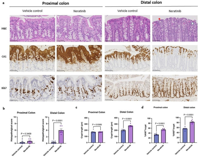
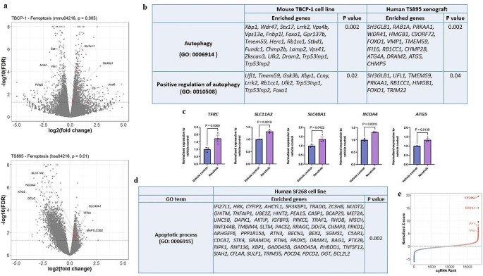
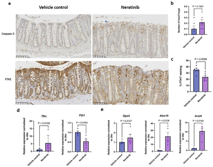
Results: The severity of colonic epithelial injury, especially degeneration of surface lining colonocytes and infiltration of immune cells, was more pronounced in the distal colon than the proximal colon. Sequencing showed that apoptotic gene signature was enriched in neratinib-treated SF268 cells while ferroptotic gene signature was enriched in neratinib-treated TBCP1 cells and T895 xenograft. However, we found that ferroptosis, but less likely apoptosis, was a potential histopathological feature underlying colonic injury in rats treated with neratinib.
Conclusion: Ferroptosis is a potential feature of neratinib-induced colonic injury and that targeting molecular machinery governing neratinib-induced ferroptosis may represent an attractive therapeutic approach to ameliorate symptoms of gut toxicity.
Keywords: Colonic injury; Ferroptosis; Gut toxicity; Neratinib.
© 2024. The Author(s).
Conflict of interest statement
The authors declare no competing interests.
Figures
Similar articles
-
Neoadjuvant neratinib promotes ferroptosis and inhibits brain metastasis in a novel syngeneic model of spontaneous HER2+ve breast cancer metastasis.Breast Cancer Res. 2019 Aug 13;21(1):94. doi: 10.1186/s13058-019-1177-1. Breast Cancer Res. 2019. PMID: 31409375 Free PMC article.
-
Preclinical and clinical evaluation through serial colonoscopic evaluation of neratinib-induced diarrhea in HER2-positive breast cancer-A pilot study.Physiol Rep. 2024 Aug;12(16):e70008. doi: 10.14814/phy2.70008. Physiol Rep. 2024. PMID: 39187401 Free PMC article. Clinical Trial.
-
Targeting neratinib-induced diarrhea with budesonide and colesevelam in a rat model.Cancer Chemother Pharmacol. 2019 Mar;83(3):531-543. doi: 10.1007/s00280-018-3756-8. Epub 2018 Dec 10. Cancer Chemother Pharmacol. 2019. PMID: 30535958
-
Neratinib, A Novel HER2-Targeted Tyrosine Kinase Inhibitor.Clin Breast Cancer. 2016 Oct;16(5):344-348. doi: 10.1016/j.clbc.2016.05.016. Epub 2016 May 29. Clin Breast Cancer. 2016. PMID: 27405796 Review.
-
Pharmacodynamics, pharmacokinetics and clinical efficacy of neratinib in HER2-positive breast cancer and breast cancer with HER2 mutations.Expert Opin Drug Metab Toxicol. 2016 Aug;12(8):947-57. doi: 10.1080/17425255.2016.1198317. Epub 2016 Jun 27. Expert Opin Drug Metab Toxicol. 2016. PMID: 27284682 Review.
References
-
- Sung H, Ferlay J, Siegel RL, Laversanne M, Soerjomataram I, Jemal A, Bray F (2021) Global Cancer statistics 2020: GLOBOCAN estimates of incidence and mortality worldwide for 36 cancers in 185 countries. CA Cancer J Clin 71(3):209–249. 10.3322/caac.21660 - PubMed
-
- Prati R, Apple SK, He J, Gornbein JA, Chang HR (2005) Histopathologic characteristics Predicting HER-2/neu amplification in breast Cancer. Breast J 11(6):433–439 - PubMed
-
- Altundag K, Bondy ML, Mirza NQ, Kau S-W, Broglio K, Hortobagyi GN, Rivera E (2007) Clinicopathologic characteristics and prognostic factors in 420 metastatic breast cancer patients with central nervous system metastasis. Cancer 110(12):2640–2647 - PubMed
-
- Gonzalez-Angulo AM, Litton JK, Broglio KR, Meric-Bernstam F, Rakkhit R, Cardoso F, Peintinger F, Hanrahan EO, Sahin A, Guray M, Larsimont D, Feoli F, Stranzl H, Buchholz TA, Valero V, Theriault R, Piccart-Gebhart M, Ravdin PM, Berry DA, Hortobagyi GN (2009) High risk of recurrence for patients with breast Cancer who have human epidermal growth factor receptor 2–Positive, node-negative tumors 1 cm or smaller. J Clin Oncol 27(34):5700–5706 - PMC - PubMed
-
- Goyette M-A, Duhamel S, Aubert L, Pelletier A, Savage P, Thibault M-P, Johnson RM, Carmeliet P, Basik M, Gaboury L, Muller WJ, Park M, Roux PP, Gratton J-P, Côté J-F (2018) The receptor tyrosine kinase AXL is required at multiple steps of the Metastatic Cascade during HER2-Positive breast Cancer Progression. Cell Rep 23(5):1476–1490 - PubMed
MeSH terms
Substances
LinkOut - more resources
Full Text Sources
Research Materials
Miscellaneous
