Review
. 2024 Aug 7;112(15):2486-2502.
doi: 10.1016/j.neuron.2024.06.014. Epub 2024 Jul 12.

Cortico-basal ganglia plasticity in motor learning

Affiliations
Review

Cortico-basal ganglia plasticity in motor learning

Richard H Roth et al. Neuron. .

Abstract

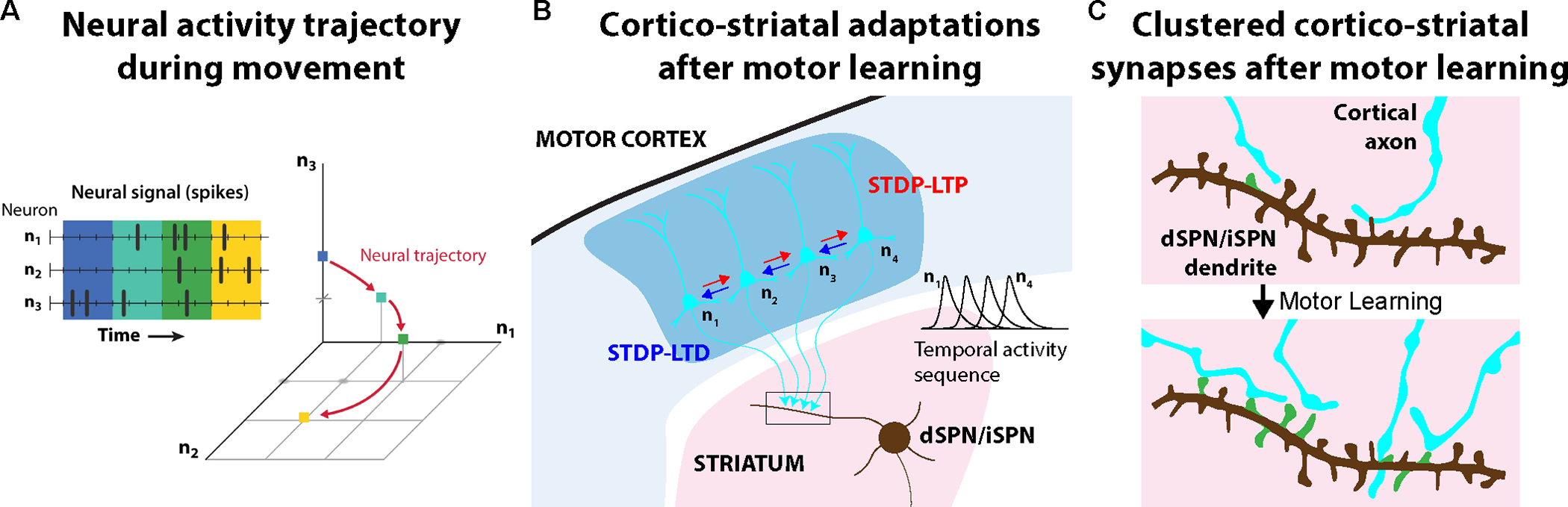
One key function of the brain is to control our body's movements, allowing us to interact with the world around us. Yet, many motor behaviors are not innate but require learning through repeated practice. Among the brain's motor regions, the cortico-basal ganglia circuit is particularly crucial for acquiring and executing motor skills, and neuronal activity in these regions is directly linked to movement parameters. Cell-type-specific adaptations of activity patterns and synaptic connectivity support the learning of new motor skills. Functionally, neuronal activity sequences become structured and associated with learned movements. On the synaptic level, specific connections become potentiated during learning through mechanisms such as long-term synaptic plasticity and dendritic spine dynamics, which are thought to mediate functional circuit plasticity. These synaptic and circuit adaptations within the cortico-basal ganglia circuitry are thus critical for motor skill acquisition, and disruptions in this plasticity can contribute to movement disorders.

Keywords: basal ganglia; circuit adaptation; dendritic spines; motor control; motor cortex; motor learning; striatum; synaptic plasticity.


Conflict of interest statement

Declaration of interests The authors declare no competing interests.

Figures

Figure 1:
Figure 1:. Brain motor circuits.
Overview of prominent brain motor circuits, including primary and secondary motor cortex (M1 and M2, blue); the basal ganglia system (red) with its input structure, the striatum, external globus pallidus (GPe) and subthalamic nucleus (STN), as well as its outputs, the internal globus pallidus (GPi) and substantia nigra pars reticulata (SNr), and the dopaminergic substantia nigra pars compacta (SNc); the thalamus (orange) with the motor thalamus (MoThal) nuclei VM and VAL as well as the parafascicular nucleus (Pf); the cerebellum (grey) with its outputs, the deep cerebellar nuclei (DCN); and the brainstem motor nuclei (green). Major projection pathways are shown by arrows.
Figure 2:
Figure 2:. Detailed view of the cortico-striatal microcircuitry.
Two classes of excitatory neurons in motor cortex – pyramidal tract (PT) and intratelencephalic (IT) neurons – innervate the striatum. While PT neurons are exclusively located in layer 5B (L5B) and send axonal collaterals towards the brainstem, spinal cord, thalamus, and striatum, IT neurons are in layers 2/3, 5A and 5B and only target the striatum as well as other cortical areas., In addition, the cortex is comprised of three major classes of inhibitory interneurons – somatostatin (SOM), parvalbumin (PV), and vasoactive intestinal peptide (VIP) expressing neurons. The striatum acts as the input nucleus of the basal ganglia system and is predominantly comprised of D1R-expressing direct pathway spiny projection neurons (dSPNs) and D2R-expressing SPNs of the indirect pathway (iSPNs). Cholinergic (ChIn) and GABAergic (including PV-, SOM-, calretinin- and Tyrosine hydroxylase-expressing) neurons make up the majority of local interneurons in the striatum. Additional input projections to cortex include thalamo-cortical and cortico-cortical projections and striatal neurons receive additional input from thalamo-striatal projections. Dopaminergic projections predominantly target the striatum but also motor cortex.
Figure 3:
Figure 3:. Common behavioral tasks used to study circuit and synaptic plasticity during motor learning in rodents.
(A) Schematic of mouse performing freely moving forelimb reaching task. (B) Mice learn to perform successful reaches over the course of 8 days of training and maintain proficiency for >30 days. (C) Reach trajectory kinematics analysis. Yellow, 30 individual reach trajectories; red, average reach trajectory. (D) Variance of reaching trajectories decreases over time of learning. (B-D adapted from Albarran et al.). (E) Schematic of mice performing head-fixed lever pushing task. (F) Mice learn to perform successful pushes over the course of training. (G) Example traces of the lever-pushing movement in one mouse on days 1 and 12 of training, showing the development of stereotyped movements. Grey, individual lever trajectories; black, average trajectory. (H) Pairwise correlation of lever-pushing trajectories for all trial pairs between adjacent days increases over the course of learning. (E-H adapted from Sheng et al.). (I) Schematic of head-fixed treadmill running in mice. (J) Mice increase running velocity over course of motor learning. Mice with and without a GRIN lens implantation exhibit comparable running adaptation. (I,J adapted from Ma et al.). (K) Example gait patterns of mice running on treadmill. (L) Running gait patterns become more structured after training. (K,L adapted from Adler et al.).
Figure 4:
Figure 4:. Circuit adaptations in motor cortex and striatum.
(A) Schematic of mouse performing head-fixed lever pushing task and task structure. (B) Activity onsets of excitatory neurons in L2/3 of motor cortex become more refined and shifted towards the beginning of movements over the course of motor learning. (C) Correlation between the neuronal activity and the learned activity pattern increases with increasing correlation between trial movement and the learned movement pattern in expert sessions but not in naïve mice. (D) A stronger correlation between population activity and movement emerges during learning. (A-D adapted from Peters et al.). (E) Average neuronal activity of dSPNs in striatum become more refined and shifted towards the beginning of movements over the course of motor learning. (F) Average neuronal activity of iSPNs in striatum become more refined and shifted towards the beginning of movements. (G,H) Movements with similar trajectories have higher correlated striatal activity following motor learning. (E-H adapted from Sheng et al.).
Figure 5:
Figure 5:. Dendritic spine plasticity in motor cortex.
(A) Schematic of mouse performing forelimb reaching task. (B) Longitudinal imaging of the same apical dendritic branches of motor cortex layer 5 neurons in Thy1-YFP mice over one-day intervals reveals spine elimination (arrows) and formation (arrowheads), and filopodia (asterisks) in trained and control mice. (A,B adapted from Xu et al.). (C) Motor learning results in a transient increase in spine density. (D) Motor learning induces an early increase in new spine additions (top) and delayed increase in spine elimination (bottom). (E) Motor learning stabilizes newly formed spines. (F) Schematic summary of spine dynamics and plasticity during motor learning. During early phases of learning new spines are formed and existing synapses are strengthened. In late phases initially potentiated synapses remain potentiated and spines formed in spatial clusters are selectively stabilized,,.
Figure 6:
Figure 6:. Common behavioral tasks used to study motor learning in primates.
(A) Schematic of monkey performing a center-out joystick reaching task (left). Reaching trajectories of initial and late sessions with clockwise and counter-clockwise curl force fields applied. Monkeys learn to adapt their trajectories to with applied force (right). (A adapted from Perich et al.). (B) Schematic of human performing a joystick reaching task (left). A divergent force field is applied such that any deviation from a straight reaching trajectory will generate a force that acts in the same direction. Humans learn to make straight movements in this task setting (right) (B adapted from Davidson et al.). (C) Motor cortex recordings are used to control neuroprosthetic devices such as moving a cursor on a screen in a center-out movement trajectory (C adapted from Wilson et al.).
Figure 7:
Figure 7:. Relationship between neural population dynamics and cortico-striatal plasticity.
(A) Neural state space representing the spiking activity from three neurons across time (colors) (A adapted from Vyas et al.). Such neural population dynamics can encode movement parameters. (B) During motor learning neurons in the motor cortex form refined temporal activity sequences that are likely mediated by synaptic plasticity mechanisms, such as spike time dependent plasticity (STDP-LTP and STDP-LTD), in cortex. (C) Cortico-striatal projections also undergo synaptic plasticity with likely post-synaptic spatial and temporal clustering, which can have an supra-linear effect on neuronal activity through local dendritic computations.

References

    1. Guo JZ, Graves AR, Guo WW, Zheng J, Lee A, Rodríguez-Gonzá Lez J, Li N, Macklin JJ, Phillips JW, Mensh BD, et al. (2015). Cortex commands the performance of skilled movement. Elife. 10.7554/eLife.10774. - DOI - PMC - PubMed
    1. Ayling OGS, Harrison TC, Boyd JD, Goroshkov A, and Murphy TH (2009). Automated light-based mapping of motor cortex by photoactivation of channelrhodopsin-2 transgenic mice. Nat. Methods 6, 219–224. 10.1038/nmeth.1303. - DOI - PubMed
    1. Ebbesen CL, and Brecht M (2017). Motor cortex - To act or not to act? Nat. Rev. Neurosci. 18. 10.1038/nrn.2017.119. - DOI - PubMed
    1. Shenoy KV, Sahani M, and Churchland MM (2013). Cortical control of arm movements: A dynamical systems perspective. Annu. Rev. Neurosci. 36. 10.1146/annurev-neuro-062111-150509. - DOI - PubMed
    1. Klaus A, Alves da Silva J, and Costa RM (2019). What, If, and When to Move: Basal Ganglia Circuits and Self-Paced Action Initiation. Annu. Rev. Neurosci. 42, annurev-neuro-072116–031033. 10.1146/annurev-neuro-072116-031033. - DOI - PubMed

LinkOut - more resources