. 2024 Aug;300(8):107552.
doi: 10.1016/j.jbc.2024.107552. Epub 2024 Jul 11.

GGA1 interacts with the endosomal Na+/H+ exchanger NHE6 governing localization to the endosome compartment

Affiliations

GGA1 interacts with the endosomal Na+/H+ exchanger NHE6 governing localization to the endosome compartment

Li Ma et al. J Biol Chem. 2024 Aug.

Abstract

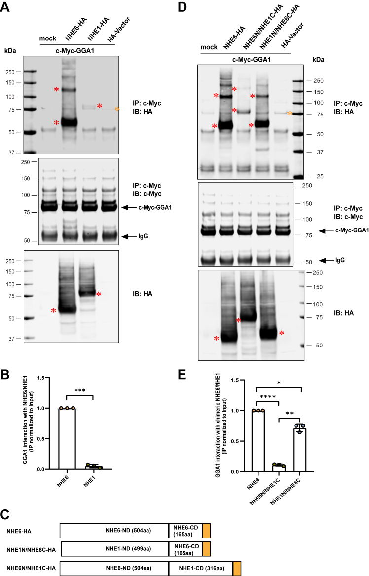
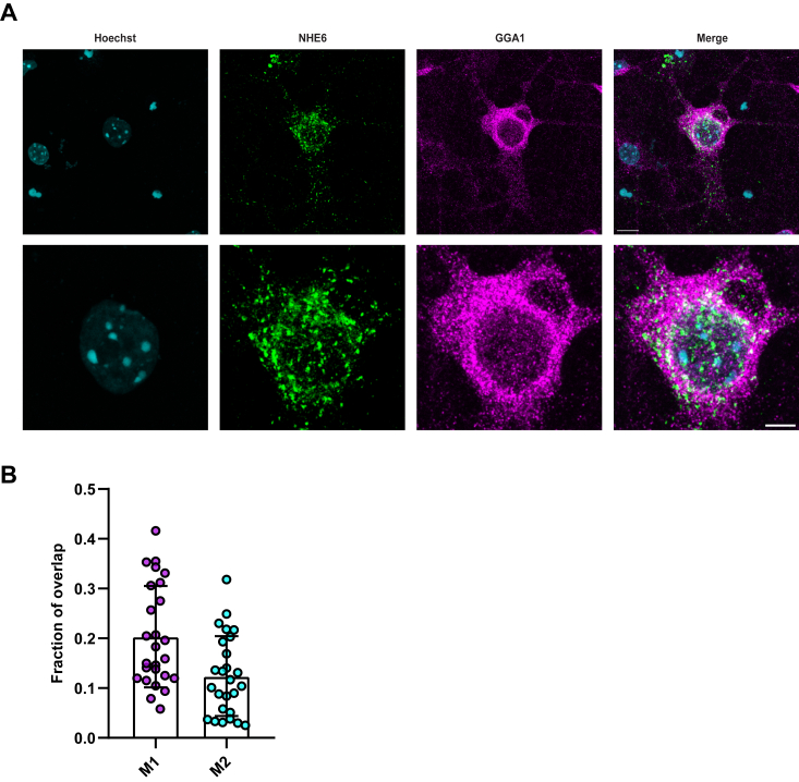
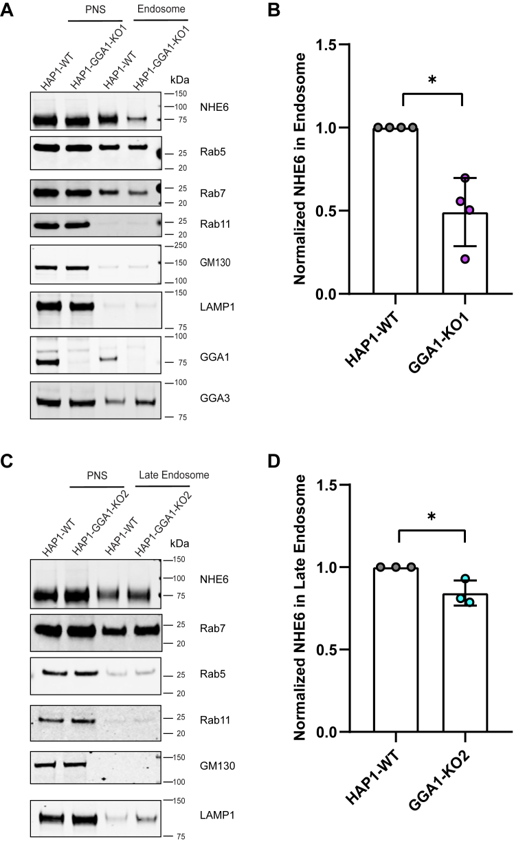
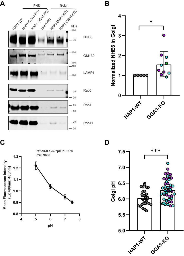
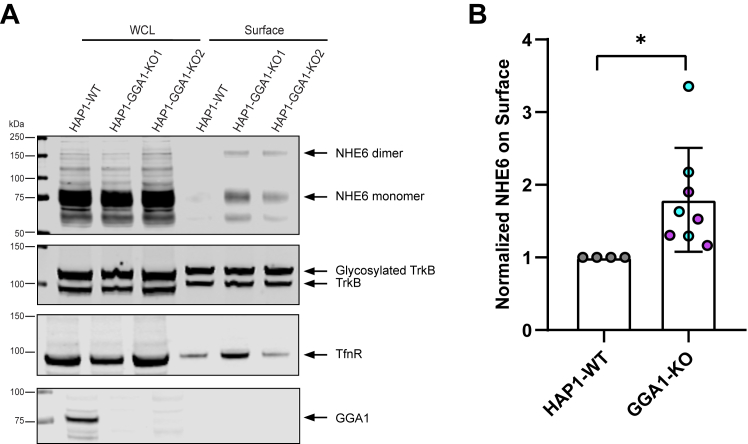
Mutations in the endosomal Na+/H+ exchanger 6 (NHE6) cause Christianson syndrome, an X-linked neurological disorder. NHE6 functions in regulation of endosome acidification and maturation in neurons. Using yeast two-hybrid screening with the NHE6 carboxyl terminus as bait, we identify Golgi-associated, gamma adaptin ear-containing, ADP-ribosylation factor (ARF) binding protein 1 (GGA1) as an interacting partner for NHE6. We corroborated the NHE6-GGA1 interaction using: coimmunoprecipitation; overexpressed constructs in mammalian cells; and coimmunoprecipitation of endogenously expressed GGA1 and NHE6 from neuroblastoma cells, as well as from the mouse brain. We demonstrate that GGA1 interacts with organellar NHEs (NHE6, NHE7, and NHE9) and that there is significantly less interaction with cell-surface localized NHEs (NHE1 and NHE5). By constructing hybrid NHE1/NHE6 exchangers, we demonstrate the cytoplasmic tail of NHE6 interacts most strongly with GGA1. We demonstrate the colocalization of NHE6 and GGA1 in cultured, primary hippocampal neurons, using super-resolution microscopy. We test the hypothesis that the interaction of NHE6 and GGA1 functions in the localization of NHE6 to the endosome compartment. Using subcellular fractionation experiments, we show that NHE6 is mislocalized in GGA1 KO cells, wherein we find less NHE6 in endosomes, but more NHE6 transport to lysosomes, and more Golgi retention of NHE6, with increased exocytosis to the surface plasma membrane. Consistent with NHE6 mislocalization, and Golgi retention, we find the intraluminal pH in Golgi to be alkalinized in GGA1-null cells. Our study demonstrates a new interaction between NHE6 and GGA1 which functions in the localization of this intracellular NHE to the endosome compartment.

Keywords: ARF binding protein 1 (GGA1); Christianson syndrome (CS); Golgi; Golgi-associated; Na+/H+ exchanger 6 (NHE6); endosome; exchangers; gamma adaptin ear containing; intracellular trafficking; lysosome.


Conflict of interest statement

Conflict of interest The authors declare that they have no conflicts of interest with the contents of this article.

Figures

Figure 1
Figure 1
Identification of GGA1 as a new NHE6 interacting partner. A, yeast cells containing bait construct pB27-NHE6 (N-LexA-NHE6-537–630, amino acids 537–630 of Slc9a6 cloned into the pB27 plasmid hgx2030v2_pB27) and prey constructs: pP6-GGA1 (N-GAL4-GGA1, clone RHC_RP_hgx2030v2_pB27_C-38 cloned into pP6) or pP6-Sept8 (N-GAL4-Sept8, clone RHC_RP_hgx2030v2_pB27_B-2 cloned into pP6). Yeast cells were obtained by mating and spotted, at the dilutions indicated, on DO-3 selective media lacking Trp, Leu, and His. Negative controls included the following empty bait or prey vectors: pB27 and pP7, or pB27+Gga1, pB27+Sept8, Slc9a6+pP7. B, cell lysates from HEK293T cells expressing mGGA1 with either NHE6-HA or HA-vector were immunoprecipitated with anti-HA antibody. The precipitates were analyzed with both anti-GGA1 and anti-NHE6 blotting. Total cell lysates (TCLs) were subjected to Western blot analysis with anti-GGA1 antibody. All samples were separated by 4 to 12% SDS–PAGE unless mentioned separately. C, cell lysates from HEK293T cells expressing c-Myc-GGA1 with either NHE6-HA or HA-vector were immunoprecipitated with anti-c-Myc antibody. The precipitates were probed with anti-NHE6 and c-Myc antibodies. TCLs were subjected to Western blot analysis with anti-HA to detect the expression of NHE6. D, Co-IP assay in SH-SY5Y neuroblastoma cells to detect interaction between endogenous NHE6 and GGA1. TCLs from SH-SY5Y cells were precipitated by anti-NHE6 antibody, with normal rabbit IgG (Santa Cruz, sc-2027) used as a control. The precipitates were subjected to Western blot analysis using anti-GGA1 and anti-NHE6 antibodies. TCLs were subjected to Western blot analysis with anti-NHE6 and anti-GGA1 to detect the expression level of endogenous NHE6 and GGA1. E, brain tissue from NHE6-null and WT males at postnatal day 0 (P0) was collected and homogenized. TCLs were precipitated by anti-NHE6 antibody, with normal rabbit IgG (Sigma-Aldrich 12–370) used as a control. The precipitates were subjected to Western blot analysis using both anti-GGA1 and anti-NHE6 antibodies. TCLs were subjected to Western blot analysis with anti-NHE6 and anti-GGA1 antibodies to detect the expression level of GGA1 and NHE6. F, TCLs from HEK293T cells expressing c-Myc-GGA1 with either HA-tagged full-length NHE6 (NHE6-FL) or cytoplasmic domain NHE6 (NHE6-CD) were immunoprecipitated with anti-c-Myc antibody. The precipitates were probed with HA and c-Myc antibodies. TCLs were subjected to Western blot analysis with anti-HA or c-Myc antibodies to detect the expression of NHE6-FL or NHE6-CD and c-Myc-GGA1. GGA1, Golgi-associated, gamma adaptin ear-containing, ARF binding protein 1; NHE6, Na+/H+ exchanger 6; Co-IP, coimmunoprecipitation; Sept8, Septin8; IgG, immunoglobulin G.
Figure 2
Figure 2
GGA1 interacts strongly with the NHE6 C-term but not with the NHE1 C-term. A, total cell lysates (TCLs) from HEK293T cells expressing c-Myc-GGA1 with either NHE6-HA or NHE1-HA were immunoprecipitated with anti-c-Myc antibody. NHE1-HA used here was mutagenesis (G720D) corrected hNHE1-HA. Control (only c-Myc-GGA1) and HA-vector (HA-vector and c-Myc-GGA1 cotransfectants) were used as control. The precipitates were probed with HA and c-Myc antibodies. TCLs were subjected to Western blot analysis with the anti-HA antibody to detect the expression of NHE6 and NHE1. Red asterisks indicate NHE6 (monomer and dimer) and NHE1 band, orange asterisk indicates a nonspecific band. B, quantification of GGA1 interaction with NHE6 and NHE1 showing a normalization of immunoprecipitated products with corresponding inputs (mean ± SD; n = 3; ∗∗∗p = 0.0003; unpaired two-tailed Student’s t test with Welch’s correction). C, domain schematics of HA-tagged NHE6, and chimeric snap constructs NHE1-N terminus+NHE6-C terminus (NHE1N/NHE6C) and NHE6-N terminus+NHE1-C terminus (NHE6N/NHE1C). D, TCLs from HEK293T cells expressing c-Myc-GGA1 with HA-tagged NHE6, NHE1N/NHE6C, and NHE6N/NHE1C were immunoprecipitated with anti-c-Myc antibody. Control (only c-Myc-GGA1) and HA-vector (HA-vector and c-Myc-GGA1 cotransfectants) were used as control. The precipitates were probed with HA and c-Myc antibodies. TCLs were subjected to Western blot analysis with anti-HA to detect the expression of NHE6 and chimeric snap constructs. Red asterisks indicate NHE6 and chimeric NHE6/NHE1 band (monomer and dimer), orange asterisk indicates a nonspecific band. E, quantification of GGA1 interaction with NHE6, NHE6N/NHE1C and NHE1N/NHE6C showing as normalization of immunoprecipitated products with corresponding inputs (mean ± SD; n = 3; ∗∗∗∗p < 0.0001 for NHE6 versus NHE6N/NHE1C; ∗p = 0.0146 for NHE6 versus NHE1N/NHE6C; ∗∗p = 0.0021 for NHE6N/NHE1C versus NHE1N/NHE6C; unpaired two-tailed Student’s t test with Welch’s correction). GGA1, Golgi-associated, gamma adaptin ear-containing, ARF binding protein 1; NHE6, Na+/H+ exchanger 6; IgG, immunoglobulin G; Ha, hemagglutinin.
Figure 3
Figure 3
Mapping of interaction regions between NHE6 and GGA1. A, structure of mGGA1 domains: VHS (aa(17–147), green), GAT (aa(171–298), purple), GAE (aa(506–627), red), Hinge (aa(299–505)), del GAE (aa(1–505)), del VHS (aa(171–635)), VHS + GAT (aa(1–298)), Hinge+GAE (aa(299–627)), GAT+Hinge (aa(171–505)), and del Hinge (del aa(299–505)). B and D, GFP-tagged GGA1 construct mGGA1-FL/VHS/GAT/Hinge/GAE (B), or GFP-tagged GGA1 construct mGGA1-FL/delVHS/delGAE/VHS+GAT/GAT+Hinge/delHinge/Hinge+GAE (D) was cotransfected with NHE6-HA in HEK293T cells. Anti-HA immunoprecipitates were analyzed with anti-GFP immunoblotting to identify GGA1 region(s) necessary for NHE6 interaction. TCLs were subjected to Western blot analysis with anti-GFP to detect the expression level of GGA1 constructs. Red asterisks indicate GGA1 FL/domains bands. C and E, quantification of GGA1 domain interaction with NHE6 showing as normalization of immunoprecipitated products with corresponding inputs. (mean ± SD; n = 3; ∗p < 0.05, ∗∗p < 0.01, ∗∗∗∗p < 0.0001; Unpaired two-tailed Student’s t test with Welch’s correction). GGA1, Golgi-associated, gamma adaptin ear-containing, ARF binding protein 1; HA, hemagglutinin; NHE6, Na+/H+ exchanger 6; TCL, total cell lysate.
Figure 4
Figure 4
NHE6 and GGA1 colocalization using confocal microscopy in mouse neurons in vitro. A, representative image of mouse primary hippocampal neurons at 14 days in vitro (14 DIV) were costained with anti-NHE6 (green) and anti-GGA1 (magenta) antibodies. Hoechst = blue. The bottom panel is a zoomed in of the top panel. The scale bar represents top panel = 10 μm; bottom panel=5 μm. B, quantification of NHE6 and GGA1 colocalization by Mander’s coefficients. M1 = fraction of NHE6 overlapping GGA1; M2 = fraction of GGA1 overlapping NHE6 (n = 26 images from three WT mice hippocampal cultures, mean ± SD). M1 and M2 are plotted as magenta and blue color, respectively. GGA1, Golgi-associated, gamma adaptin ear-containing, ARF binding protein 1; NHE6, Na+/H+ exchanger 6.
Figure 5
Figure 5
Colocalization of NHE6 and GGA1 by 5x expansion microscopy imaging in primary neurons in vitro. Colocalization of GGA1 and NHE6 with respect to Golgi in rat hippocampal neurons: (A) shows a single frame of 5× expanded rat hippocampal neuron on DIV 14 costained for nucleus (blue), Giantin (cyan), GGA1 (red), and NHE6 (green) from a z-stack acquired using confocal microscopy. Corrected scale bar represents ∼2.27 μm. The intensities were adjusted for visualization. B, enlarged image from (A) shown in yellow box. The corrected scale bar represents ∼0.68 μm. C, shows the 3D reconstruction of the z-stack using IMARIS surface tools for the nucleus (white), and Giantin (brown) and spot classification for GGA1 (green, yellow, or cyan), and NHE6 (red, pink, or blue). The spots are classified based on the colocalization of a GGA1 or NHE6 with the other stain. The corrected scale bar represents ∼2.27 μm. D, enlarged image from (C) shown in red box. The corrected scale bar represents ∼0.68 μm. E, colocalization fraction of GGA1 or NHE6 analyzed using IMARIS software. M1: fraction of GGA1 colocalizing with NHE6; M1G: fraction of colocalizing GGA1 with NHE6 inside Giantin; M2: fraction of NHE6 colocalizing with GGA1; M2G: fraction of colocalizing NHE6 with GGA1 inside Giantin. The color code for (C and D) are described to the right of image (n = 14 cells from four unique neuronal cultures). The corrected scale bar refers to scale correction after accounting for physical expansion of the cells, based on Fig. S5A. GGA1, Golgi-associated, gamma adaptin ear-containing, ARF binding protein 1; NHE6, Na+/H+ exchanger 6.
Figure 6
Figure 6
Loss of GGA1 leads to less NHE6 in endosomes. A, Western blot of NHE6 protein in early endosome fractionation in HAP1 GGA1 KO line 1 and WT cells using the following markers: Rab5 (early endosome), Rab7 (late endosome), Rab11 (recycling endosome), LAMP1 (lysosome), and GM130 (Golgi). PNS = post nuclear supernatant. B, quantification of NHE6 protein levels in endosome fractions in HAP1 GGA1-KO line 1 and WT cells. The normalization was performed as follows: NHE6 in endosomes was first normalized to NHE6 in PNS (termed A); Rab5 and Rab7 in endosomes was normalized to Rab5 and Rab7 in PNS (termed B); normalized NHE6 in endosomes was then calculated as A divided by B. (mean ± SD; n = 4; ∗p = 0.0158, Unpaired two-tailed Student’s t test with Welch’s correction). C, Western blot of NHE6 protein in late endosome fractionation in HAP1 GGA1 KO line 2 and WT cells using the following markers: Rab5 (early endosome), Rab7 (late endosome), Rab11 (recycling endosome), LAMP1 (lysosome), and GM130 (Golgi). PNS = post nuclear supernatant. D, quantification of NHE6 in late endosome fractionation in HAP1 GGA1KO line 2 and WT cells. For normalization: NHE6 in late endosomes was first normalized to NHE6 in PNS (termed A); Rab7 in late endosomes was normalized to Rab7 in PNS (termed B); normalized NHE6 in late endosomes was then calculated as A divided by B. (mean ± SD; n = 3; ∗p = 0.0237, Unpaired two-tailed Student’s t test). GGA1, Golgi-associated, gamma adaptin ear-containing, ARF binding protein 1; NHE6, Na+/H+ exchanger 6.
Figure 7
Figure 7
Loss of GGA1 leads to greater NHE6 in lysosomes but does not affect lysosome pH. A, Western blot of NHE6 protein in lysosome fractionation in HAP1 GGA1 KO and WT cells using the following markers: LAMP1 (lysosome), Rab5 (early endosome), Rab7 (late endosome), Rab11 (recycling endosome), GM130 (Golgi). PNS=post nuclear supernatant. B, quantification of NHE6 in lysosome fractionation in HAP1 GGA1 KO and WT cells. The normalization was performed as follows: NHE6 in lysosome was first normalized to NHE6 in PNS (termed A); LAMP1 in lysosome was normalized to LAMP1 in PNS (termed B); normalized NHE6 in lysosome was then calculated as A divided by B. (mean ± SD; n = 3; ∗∗∗p = 0.0002, Unpaired two-tailed Student’s t test with Welch’s correction). GGA1-KO1 and KO2 are presented as magenta and blue color, respectively. C, pH calibration curve graph for lysosome pH measurement by LysoSensor Yellow/Blue DND-160 after 1 min incubation. D, quantification of lysosomal pH in HAP1 GGA1 KO and WT cells. (mean ± SD; n = 12 replicates; ns=not significant; Unpaired two-tailed Student’s t test with Welch’s correction) GGA1-KO1 and KO2 are presented as magenta and blue color, respectively. GGA1, Golgi-associated, gamma adaptin ear-containing, ARF binding protein 1; NHE6, Na+/H+ exchanger 6.
Figure 8
Figure 8
Loss of GGA1 leads to more NHE6 in Golgi and alkalinization of Golgi pH. A, Western blot of NHE6 protein in Golgi fractionation from HAP1 GGA1 KO and WT cells using the following markers: GM130 (Golgi), LAMP1 (lysosome), Rab5 (early endosome), Rab7 (late endosome), and Rab11 (recycling endosome). PNS = post-nuclear supernatant. B, quantification of NHE6 in Golgi fractionation in HAP1 GGA1 KO and WT cells. The normalization was performed as follows: NHE6 in Golgi was first normalized to NHE6 in the PNS (termed A); GM130 in Golgi was normalized to GM130 in PNS (termed B); normalized NHE6 in Golgi was then calculated as A divided by B. (mean ± SD; n = 5; GGA1-KO1 and KO2 are presented as magenta and blue color, respectively; ∗p = 0.0207, Unpaired two-tailed Student’s t test with Welch’s correction). GGA1-KO1 and KO2 are presented as magenta and blue color, respectively. C, graph of the Golgi pH calibration curve in HAP1 GGA1 KO and WT cells using TGN38-pHluorin construct. D, quantification of Golgi pH in HAP1 GGA1 KO and WT cells. (mean ± SD; WT n = 27, GGA1 KO1 n = 22, GGA1 KO2 n = 24 cells; ∗∗∗p = 0.0005, Unpaired two-tailed Student’s t test with Welch’s correction). GGA1-KO1 and KO2 are presented as magenta and blue color, respectively. GGA1, Golgi-associated, gamma adaptin ear-containing, ARF binding protein 1; NHE6, Na+/H+ exchanger 6; TGN, trans-Golgi network.
Figure 9
Figure 9
Loss of GGA1 leads to more NHE6 on plasma membrane. A, whole cell lysate (WCL) and biotinylated surface were separated by Western blot and detected by immunoblotting with anti-NHE6, transferrin receptor (TfnR), tropomyosin receptor kinase B (TrkB), and GGA1 antibodies. TfnR and TrkB are shown to demonstrate the quality of the cell surface labeling and fractionation; whereas GGA1, an intracellular protein, is not detected in the cell surface fraction. B, quantification of NHE6 on plasma membrane in HAP1 GGA1 KO and WT cells. The normalization was performed as follows: NHE6 on the surface was first normalized to NHE6 in WCL (termed A); the plasma membrane protein TfnR on the surface was normalized to TfnR in WCL (termed B); normalized NHE6 on the surface was then calculated as A divided by B. (mean ± SD; n = 4 for each line, GGA1-KO1 and KO2 are presented as magenta and blue color, respectively; ∗p = 0.0163, Unpaired two-tailed Student’s t test with Welch’s correction). GGA1-KO1 and KO2 are presented as magenta and blue color, respectively. GGA1, Golgi-associated, gamma adaptin ear-containing, ARF binding protein 1; NHE6, Na+/H+ exchanger 6.

Update of

References

    1. Christianson A.L., Stevenson R.E., van der Meyden C.H., Pelser J., Theron F.W., van Rensburg P.L., et al. X linked severe mental retardation, craniofacial dysmorphology, epilepsy, ophthalmoplegia, and cerebellar atrophy in a large South African kindred is localised to Xq24-q27. J. Med. Genet. 1999;36:759–766. - PMC - PubMed
    1. Gilfillan G.D., Selmer K.K., Roxrud I., Smith R., Kyllerman M., Eiklid K., et al. SLC9A6 mutations cause X-linked mental retardation, microcephaly, epilepsy, and ataxia, a phenotype mimicking Angelman syndrome. Am. J. Hum. Genet. 2008;82:1003–1010. - PMC - PubMed
    1. Pescosolido M.F., Stein D.M., Schmidt M., El Achkar C.M., Sabbagh M., Rogg J.M., et al. Genetic and phenotypic diversity of NHE6 mutations in Christianson syndrome. Ann. Neurol. 2014;76:581–593. - PMC - PubMed
    1. Ouyang Q., Lizarraga S.B., Schmidt M., Yang U., Gong J., Ellisor D., et al. Christianson syndrome protein NHE6 modulates TrkB endosomal signaling required for neuronal circuit development. Neuron. 2013;80:97–112. - PMC - PubMed
    1. Lizarraga S.B., Ma L., Maguire A.M., van Dyck L.I., Wu Q., Ouyang Q., et al. Human neurons from Christianson syndrome iPSCs reveal mutation-specific responses to rescue strategies. Sci. Transl. Med. 2021;13 - PMC - PubMed

Substances

LinkOut - more resources