. 2024 Jul 13;14(1):16203.
doi: 10.1038/s41598-024-67235-y.

Building on the clinical applicability of ctDNA analysis in non-metastatic pancreatic ductal adenocarcinoma

Affiliations

Building on the clinical applicability of ctDNA analysis in non-metastatic pancreatic ductal adenocarcinoma

Ibone Labiano et al. Sci Rep. .

Abstract

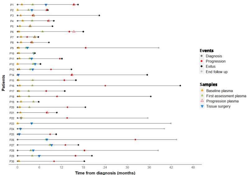
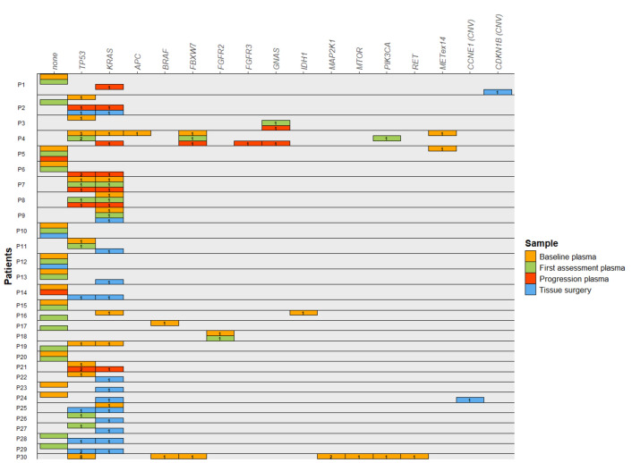
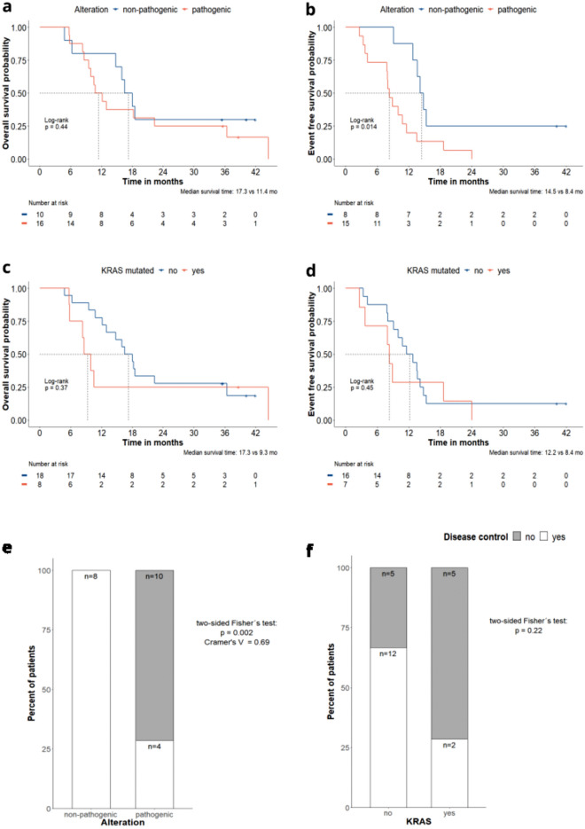
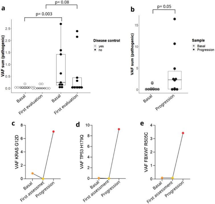
Pancreatic ductal adenocarcinoma represents one of the solid tumors showing the worst prognosis worldwide, with a high recurrence rate after adjuvant or neoadjuvant therapy. Circulating tumor DNA analysis raised as a promising non-invasive tool to characterize tumor genomics and to assess treatment response. In this study, surgical tumor tissue and sequential blood samples were analyzed by next-generation sequencing and were correlated with clinical and pathological characteristics. Thirty resectable/borderline pancreatic ductal adenocarcinoma patients treated at the Hospital Universitario de Navarra were included. Circulating tumoral DNA sequencing identified pathogenic variants in KRAS and TP53, and in other cancer-associated genes. Pathogenic variants at diagnosis were detected in patients with a poorer outcome, and were correlated with response to neoadjuvant therapy in borderline pancreatic ductal adneocarcinoma patients. Higher variant allele frequency at diagnosis was associated with worse prognosis, and thesum of variant allele frequency was greater in samples at progression. Our results build on the potential value of circulating tumor DNA for non-metastatic pancreatic ductal adenocarcinoma patients, by complementing tissue genetic information and as a non-invasive tool for treatment decision. Confirmatory studies are needed to corroborate these findings.

Keywords: Biomarkers; Gastrointestinal neoplasms; Genomics; Liquid biopsy; Precision medicine.


Conflict of interest statement

IL, AEH, SM, IGB, DGS, EM, DG and AV declare no conflict of interest; MA has been involved as a consultant for advisory roles with Amgen, BMS, MSD, Lilly and Servier; HA has been involved as a consultant for advisory roles from Astra Zeneca and for trial coordination from Ferrer Farma; NC: has received speaker honoraria from Roche and Pierre Fabre; AL has received speaker honoraria from Pierre-Fabre; APG has received speaker honoraria from Merck Sharp; GAA has received speaker honoraria from ThermoFisher and Roche; IHG has received speaker honoraria from Astra Zeneca; VA has been involved as a consultant for advisory roles and received speaker honoraria from MSD, Bristol, Lilly, Astra-Zeneca and Pierre-Fabre. RV has been involved as a consultant for advisory roles with Servier, Roche and Merck Sharp and has received speaker honoraria from Roche, Amgen, Merck Sharp and Dohme, Astra Zeneca.

Figures

Figure 1
Figure 1
Clinical events and sample collection during follow-up. Horizontal lines represent the time frame from diagnosis to the end of the follow-up for each patient. Colored diamonds represent clinical events including diagnosis (grey), progression (red), exitus (black) and end of follow-up (grey). Colored triangles represent samples collected from each patient, tissue-surgery (blue) and plasma samples at different time points including, baseline plasma (orange), first tumor assessment plasma (green) and progression plasma (red).
Figure 2
Figure 2
Summary of the pathogenic genomic alterations found in plasma and tissue samples from all patients. Horizontal boxes represent one patient, with one sub-line for each of the samples available for each patient. Colored rectangles represent the gene in which a genetic alteration was identified in each sample, including baseline plasma (orange), first tumor assessment plasma (green), progression plasma (red) and tissue surgery (blue). The label in each rectangle depicts the number of alterations observed for each gene.
Figure 3
Figure 3
Analyses based on the alterations identified in the baseline plasma sample. Kaplan-Meier analysis comparing patients showing pathogenic alterations (red) vs non-pathogenic alterations (i.e., no genetic alteration or alterations identify as non-pathogenic) (blue), (a) overall survival (b) event free survival. Kaplan-Meier analysis comparing patients showing KRAS mutations (red) vs no KRAS mutations (blue), (c) overall survival (d) event free survival. +Represents the censored patients. (e) Comparison of the proportion of patients with disease control (white) vs no control (grey) in those with pathogenic alterations vs with non-pathogenic alterations (i.e., no genetic alteration or alterations identified as non-pathogenic). (f) Comparison of the proportion of patients with disease control (white) vs no control (grey) in those with KRAS mutation vs no KRAS mutation.
Figure 4
Figure 4
Variant allele frequency changes. Data are represented as with median and interquartile range. (a) Comparison in the VAF sum of pathogenic alterations between those with disease control at first tumor assessment (white) and those without (black) in both baseline plasma sample and first tumor assessment plasma sample. (b) Comparison in the VAF sum of pathogenic alterations between baseline plasma sample and progression plasma sample. (c-e) Dynamics of the VAF in some subjects in which the same alteration was observed in baseline, first tumor assessment and progression plasma samples. All comparisons were performed by Mann-Whitney U test.

References

    1. Taieb J, Abdallah R. How I treat pancreatic cancer. ESMO Open. 2020;4(Suppl 2):e000818. doi: 10.1136/esmoopen-2020-000818. - DOI - PMC - PubMed
    1. Wood LD, Canto MI, Jaffee EM, Simeone DM. Pancreatic cancer: Pathogenesis, screening, diagnosis, and treatment. Gastroenterology. 2022;163(2):386–402.e1. doi: 10.1053/j.gastro.2022.03.056. - DOI - PMC - PubMed
    1. Sung H, et al. Global cancer statistics 2020: GLOBOCAN estimates of incidence and mortality worldwide for 36 cancers in 185 countries. CA Cancer J. Clin. 2021;71(3):209–249. doi: 10.3322/caac.21660. - DOI - PubMed
    1. Ferlay J, Partensky C, Bray F. More deaths from pancreatic cancer than breast cancer in the EU by 2017. Acta Oncol. 2016;55(9–10):1158–1160. doi: 10.1080/0284186X.2016.1197419. - DOI - PubMed
    1. Neoptolemos JP, et al. Comparison of adjuvant gemcitabine and capecitabine with gemcitabine monotherapy in patients with resected pancreatic cancer (ESPAC-4): A multicentre, open-label, randomised, phase 3 trial. Lancet. 2017;389(10073):1011–1024. doi: 10.1016/S0140-6736(16)32409-6. - DOI - PubMed

MeSH terms