. 2024 Jul 23;43(7):114479.
doi: 10.1016/j.celrep.2024.114479. Epub 2024 Jul 13.

Multiple transatlantic incursions of highly pathogenic avian influenza clade 2.3.4.4b A(H5N5) virus into North America and spillover to mammals

Affiliations

Multiple transatlantic incursions of highly pathogenic avian influenza clade 2.3.4.4b A(H5N5) virus into North America and spillover to mammals

Cassidy N G Erdelyan et al. Cell Rep. .

Abstract

Highly pathogenic avian influenza (HPAI) viruses have spread at an unprecedented scale, leading to mass mortalities in birds and mammals. In 2023, a transatlantic incursion of HPAI A(H5N5) viruses into North America was detected, followed shortly thereafter by a mammalian detection. As these A(H5N5) viruses were similar to contemporary viruses described in Eurasia, the transatlantic spread of A(H5N5) viruses was most likely facilitated by pelagic seabirds. Some of the Canadian A(H5N5) viruses from birds and mammals possessed the PB2-E627K substitution known to facilitate adaptation to mammals. Ferrets inoculated with A(H5N5) viruses showed rapid, severe disease onset, with some evidence of direct contact transmission. However, these viruses have maintained receptor binding traits of avian influenza viruses and were susceptible to oseltamivir and zanamivir. Understanding the factors influencing the virulence and transmission of A(H5N5) in migratory birds and mammals is critical to minimize impacts on wildlife and public health.

Keywords: A(H5N5); CP: Microbiology; HPAI; Sable Island; antiviral susceptibility; avian influenza; clade 2.3.4.4b; contact transmission; ferret model; wildlife transmission.


Conflict of interest statement

Declaration of interests The authors declare no competing interests.

Figures

Figure 1.
Figure 1.. A(H5N5) cases in the Atlantic provinces of Canada (n = 41)
(A) Locations where animals were collected. (B) Type of wild bird and mammalian species that virus was detected in.
Figure 2.
Figure 2.. Bayesian analyses of viral WGS using BEAST to examine the relationship of A(H5N5) viruses
(A) Bayesian timescaled maximum clade credibility tree. Inset contains a tetrameric model of NA with the region containing the 22-amino acid stalk deletion highlighted in red; PDB: 6CRD. (B) Chord diagram showing Bayesian host transition between species in Canadian A(H5N5) isolates; internal coloring matching the outer ring indicates the source species. Outer rings not matching internal coloring indicate sink species. Chords are statistically supported (Bayes factor ≥3.0). (C) Bayesian phylogeographic reconstruction of A(H5N5) dissemination (2020–2023) between European countries and the introduction of the virus into North America.
Figure 3.
Figure 3.. Pathogenicity and transmission potential of the A(H5N5) viruses in ferrets
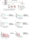
(A) Experimental design of ferret pathogenesis and transmission of A/American_Crow/PEI/FAV-0035-6/2023 (H5N5, Crow) and A/Raccoon/PEI/FAV-0193-1/2023 (H5N5, Raccoon) viruses. At 0 dpi, 9 ferrets were inoculated with 106 median egg infectious doseunits of each A(H5N5) virus. Three inoculated ferrets (Donor) were individually co-housed with 3 naive contact ferrets (Direct contact) on 1 dpi. Six ferrets were euthanized at 4 dpi for viral titration in tissues and pathology (n = 3 each). All ferrets were monitored for clinical signs of infection until day 14. Nasal wash samples were collected from both infected and direct contact ferrets at the indicated time points for virus titration. Serum samples were collected from all surviving ferrets at 25 dpi for seroconversion assays. (B–E) Survival curve (B), (C) temperature change of inoculated ferrets (values are the average ± SE for each group), (D) weight changes (ferret weight values are the average ± SE for each group), and (E) clinical scores of inoculated ferrets (n = 3 per virus). (F) Infectious viral titers from collected tissues (n = 3 ferrets). (G) Infectious viral titers from nasal washes (mean virus titer ± SD). Symbols represent each individual animal’s titer. Dashed lines indicate the lower limit of virus titer detection (1.0 log10 TCID50/mL).
Figure 4.
Figure 4.. Ferrets infected with the A/Raccoon/PEI/FAV-0193-1/2023 (H5N5) virus
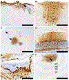
(A–F) Histopathology and immunohistochemistry showing widespread CNS infection in several locations and cell types, including (A) meninges and submeningeal neuropil, (B) cortical astrocytes and neurons, (C) perivascular neuropil in cortex, (D) Purkinje cells and glial cells in cerebellum, (E) ventricular ependyma and subependymal cells (arrow) in midbrain, and (F) infection extending from the olfactory ventricle (arrow) into surrounding layear of the olfactory bulb. Granule cell layer (gcl); mitral cell layer (mcl); external plexiform layer (epl); glomerular layer (gl). Scale bars: (A), (B), and (D) 200 μm; (C) and (F) 1 mm; (E) 100 μm.
Figure 5.
Figure 5.. Ferrets infected with the A/American_Crow/PEI/FAV-0035-6/2023 virus
(A–D) Histopathology and immunohistochemistry showing widespread CNS infection in several locations and cell types, including (A) meninges (arrow) and neuropil, (B) glial cells and some neurons in the cortex, (C) ependymal cells (arrow), and (D) choroid plexus (arrow) and periventricular ependyma/subependyma. Scale bars: (A) 200 μm; (B) and (D) 100 μm; (C) 50 μm.
Figure 6.
Figure 6.. Migration pattern of black-legged kittiwakes and northern fulmar from Norwegian colonies
(A) Black-legged kittiwake, 2021–2022; Anda, orange; Runde and Ålesund, blue; Sklinna, green. (B) Northern fulmar, 2011–2022; Jarsteinen, purple.

References

    1. Kosik I, and Yewdell JW (2019). Influenza Hemagglutinin and Neuraminidase: Yin−Yang Proteins Coevolving to Thwart Immunity. Viruses 11, 346. - PMC - PubMed
    1. Dhingra MS, Artois J, Dellicour S, Lemey P, Dauphin G, Von Dobschuetz S, Van Boeckel TP, Castellan DM, Morzaria S, and Gilbert M (2018). Geographical and Historical Patterns in the Emergences of Novel Highly Pathogenic Avian Influenza (HPAI) H5 and H7 Viruses in Poultry. Front. Vet. Sci 5, 84. 10.3389/fvets.2018.00084. - DOI - PMC - PubMed
    1. Webster RG, Bean WJ, Gorman OT, Chambers TM, and Kawaoka Y (1992). Evolution and ecology of influenza A viruses. Viruses 56, 152–179. 10.1128/mr.56.1.152-179.1992. - DOI - PMC - PubMed
    1. Gilbert M, Xiao X, Domenech J, Lubroth J, Martin V, and Slingenbergh J (2006). Anatidae Migration in the Western Palearctic and Spread of Highly Pathogenic Avian Influenza H5N1 Virus. Emerg. Infect. Dis 12, 1650–1656. 12. 10.3201/eid1211.060223. - DOI - PMC - PubMed
    1. Kawaoka Y, Chambers TM, Sladen WL, and Webster RG (1988). Is the gene pool of influenza viruses in shorebirds and gulls different from that in wild ducks? Virology 163, 247–250. 10.1016/0042-6822(88)90260-7. - DOI - PubMed

Publication types