. 2024 Jul 15;13(1):25.
doi: 10.1038/s41389-024-00527-0.

YAP/TAZ interacts with RBM39 to confer resistance against indisulam

Affiliations

YAP/TAZ interacts with RBM39 to confer resistance against indisulam

Toshinori Ando et al. Oncogenesis. .

Abstract

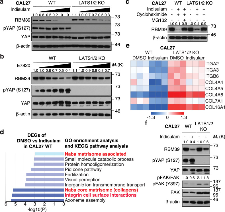
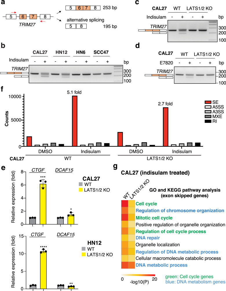
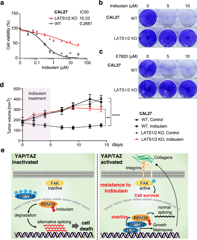
The Hippo pathway and its downstream effectors, Yes-associated protein/transcriptional coactivator with PDZ-binding motif (YAP/TAZ), are essential for cell growth and organ development. Emerging evidence revealed that the Hippo pathway and YAP/TAZ are frequently dysregulated by multiple genetic alterations in solid cancers including head and neck squamous cell carcinoma (HNSCC); however, the YAP/TAZ-nuclear interactome remains unclear. RNA-binding motif protein 39 (RBM39) enhances transcriptional activity of several transcription factors and also regulates mRNA splicing. Indisulam degrading RBM39 induces alternative splicing, leading to cell death. However, clinical trials of indisulam have failed to show effectiveness. Therefore, clarifying the resistance mechanism against splicing inhibitors is urgently required. In this study, we identified RBM39 as a novel YAP/TAZ-interacting molecule by proteome analysis. RBM39 promoted YAP/TAZ transcriptional activity. We further elucidated that indisulam reduces RBM39/YAP/TAZ-mediated integrin or collagen expression, thereby inactivating focal adhesion kinase (FAK) important for cell survival. Moreover, indisulam also induced alternative splicing of cell cycle- or DNA metabolism-related genes. YAP/TAZ hyperactivation delayed indisulam-induced RBM39 degradation, which restored the integrin/collagen expression to activate FAK, and alternative splicing, thereby conferring resistance against indisulam in vitro and in vivo. Our findings may aid to develop a novel cancer therapy focusing on YAP/TAZ/RBM39 interaction.


Conflict of interest statement

The authors declare no competing interests.

Figures

Fig. 1
Fig. 1. RBM39 promotes YAP/TAZ transcriptional activity and YAP/TAZ active cells are resistant to indisulam.
a Schematic representation of RBM39 identification. Lysate was co-immunoprecipitated with anti-YAP antibody, then analyzed by proteome analysis. The result was compared with the BioGRID database. b Western blot of HEK293A cells overexpressing Myc-YAP and HA-RBM39. Lysate was co-immunoprecipitated with anti-Myc antibody. c Immunofluorescence of proximity ligation assay in LATS1/2 KO CAL27 cells. The red signal suggests the protein interaction. The antibodies against YAP and RBM39 were used. d Relative mRNA expression in HEK293A cells overexpressing vector or RBM39. Cells were transfected with the vectors, then treated with LATS-IN-1 (10 μM) for 24 h. (N = 3). e The DEPMAP shows the top 20 gene expression conferring resistance to indisulam (Pearson ratio is gene expression vs drug sensitivity area under the curve (AUC)). YAP1 and WWTR1 are highlighted in red, and YAP/TAZ-targeted genes in green. f Western blot of HNSCC cell lines. Cells were treated with indisulam at 0, 0.01, 0.05, 0.1, 0.5, and 1 μM for 24 h. g Cell viability of HNSCC cell lines. (N = 5). h Crystal violet staining of viable HNSCC cells. ANOVA with Tukey–Kramer post hoc test was used. Mean ± SEM (d); ****, P < 0.0001; ***, P < 0.001; **, P < 0.01. Protein level of RBM39 was compared to without indisulam treatment (f).
Fig. 2
Fig. 2. YAP/TAZ delay RBM39 degradation and promote integrin, collagen, and FAK activation to be resistant to indisulam.
a Western blot of WT and LATS1/2 KO CAL27 cells treated with indisulam at 0, 0.01, 0.05, 0.1, 0.5, and 1 μM for 24 h. b Western blot treated with E7820. c Western blot of WT and LATS1/2 KO CAL27 cells treated with indisulam at 1 μM, cycloheximide at 200 μg/mL, and MG132 at 10 μM for 8 h. d GO and KEGG pathway enrichment analysis of DEGs in WT CAL27 cells treated with DMSO or indisulam. e Heatmap of integrins and collagens expression. f Western blot of WT and LATS1/2 KO CAL27 cells. Cells were treated with indisulam at 1 μM for 24 h. Protein level of RBM39 was compared to WT CAL27 cells without indisulam, E7820, cycloheximide, or MG132 treatment (ac, f).
Fig. 3
Fig. 3. LATS1/2 KO-driven YAP/TAZ activation suppresses indisulam-induced alternative splicing.
a Schematic representation of the PCR products of TRIM27 detecting alternative splicing. b Alternative splicing of TRIM27 in HNSCC cell lines. c Alternative splicing of TRIM27 in WT and LATS1/2 KO CAL27 cells treated with indisulam. (d) Alternative splicing treated with E7820. (e) Relative mRNA expression of CTGF and DCAF15 in WT and LATS1/2 KO CAL27 cells (N = 3). (f) Comprehensive splicing analysis of DMSO or indisulam-treated WT and LATS1/2 KO CAL27 cells. SE skipped exon, A5SS alternative 3’ splice site, A3SS alternative 5’ splice site, MXE mutually excluded exon, RI retained intron. (g) GO and KEGG pathway enrichment analysis of the SE genes. Cell cycle-related are highlighted in green, and DNA-metabolism related are in blue. Student’s t-test was used. Mean ± SEM (e); ****, P < 0.0001; ***, P < 0.001; **, P < 0.01.
Fig. 4
Fig. 4. LATS1/2 KO-driven YAP/TAZ activation confers resistance against indisulam in vitro and in vivo.
a Cell viability of WT and LATS1/2 KO CAL27 cells. (N = 3). b Crystal violet staining of viable WT and LATS1/2 KO CAL27 cells treated with indisulam. c Crystal violet staining treated with E7820. d Average growth curves for WT and LATS1/2 KO CAL27 cells transplanted into athymic nude mice. The tumors were treated with indisulam for 6 days (N = 10 per group). e Summary of the mechanism. ANOVA with Tukey–Kramer post hoc test was used. Mean ± SEM (d); ****, P < 0.0001; **, P < 0.01.

References

    1. Yu FX, Zhao B, Guan KL. Hippo pathway in organ size control, tissue homeostasis, and cancer. Cell. 2015;163:811–28. doi: 10.1016/j.cell.2015.10.044. - DOI - PMC - PubMed
    1. Ando T, Okamoto K, Shintani T, Yanamoto S, Miyauchi M, Gutkind JS, et al. Integrating genetic alterations and the Hippo Pathway in head and neck squamous cell carcinoma for future precision medicine. J Pers Med. 2022;12:1544. doi: 10.3390/jpm12101544. - DOI - PMC - PubMed
    1. Martin D, Degese MS, Vitale-Cross L, Iglesias-Bartolome R, Valera JLC, Wang Z, et al. Assembly and activation of the Hippo signalome by FAT1 tumor suppressor. Nat Commun. 2018;9:2372. doi: 10.1038/s41467-018-04590-1. - DOI - PMC - PubMed
    1. Ando T, Arang N, Wang Z, Costea DE, Feng X, Goto Y, et al. EGFR regulates the Hippo pathway by promoting the tyrosine phosphorylation of MOB1. Commun Biol. 2021;4:1237. doi: 10.1038/s42003-021-02744-4. - DOI - PMC - PubMed
    1. Kapoor A, Yao W, Ying H, Hua S, Liewen A, Wang Q, et al. Yap1 activation enables bypass of oncogenic Kras addiction in pancreatic cancer. Cell. 2014;158:185–97. doi: 10.1016/j.cell.2014.06.003. - DOI - PMC - PubMed