. 2024 Jul 15;8(1):21.
doi: 10.1186/s41824-024-00209-5.

FDG altered biodistribution in white adipose tissue, a rare entity: case report and review of the literature

Affiliations

FDG altered biodistribution in white adipose tissue, a rare entity: case report and review of the literature

William Y Raynor et al. EJNMMI Rep. .

Abstract

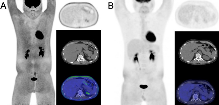
Purpose: Altered 18F-fluorodeoxyglucose (FDG) biodistribution due to patient factors such as exercise and inadequate fasting are well established causes of limited diagnostic efficacy. In addition, medications such as G-CSF are known to affect uptake of FDG by bone marrow and spleen. In this study, we present a case of increased white adipose uptake in a pediatric lymphoma patient who recently received high dose dexamethasone and review the relevant literature regarding this rare and poorly understood pattern of altered FDG biodistribution.

Methods: A 14-year-old male patient diagnosed with B-cell lymphoblastic lymphoma underwent FDG-PET/CT for restaging shortly after completing an induction chemotherapy regimen. Images revealed diffuse FDG uptake localizing to white adipose tissue, attributed to the 29-day course of dexamethasone which was completed two days prior. A diagnostically adequate study with relative normalization of FDG biodistribution was obtained seven days later.

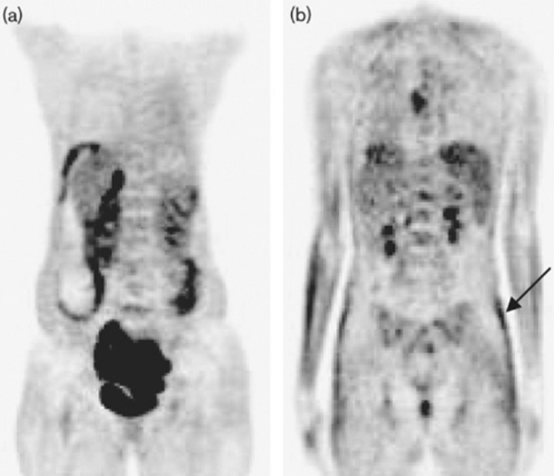
Results: In our review of the literature, diffuse FDG uptake by white fat is a rare occurrence and has only been reported by a few case reports and early observational studies. In addition to patients receiving corticosteroids, other cases of medication-induced adipose remodeling such as patients receiving highly active antiretroviral therapy have been documented with similar patterns of increased white adipose tissue activity.

Conclusion: Corticosteroid-induced white fat uptake of FDG is a rare phenomenon that can limit diagnostic accuracy of FDG-PET/CT and necessitate repeat imaging. Current evidence suggests that a wait period of at least one week after discontinuation of corticosteroids is sufficient to allow for decreased white fat uptake and increased diagnostic accuracy.

Keywords: Adipogenesis; Corticosteroids; Dexamethasone; Glucocorticoids; Lymphoma; PET/CT.


Conflict of interest statement

The authors have no relevant financial or non-financial interests to disclose.

Figures

Fig. 1
Fig. 1
FDG-PET/CT images for restaging approximately one month after initial baseline imaging. Obtained two days after the patient finished a 29-day regimen of high-dose dexamethasone, the initial study (A) clearly shows diffuse FDG uptake in white adipose tissue, as seen on the maximum intensity projection (MIP), axial PET, and fused PET/CT imaging, limiting diagnostic utility. Repeat FDG-PET/CT performed 7 days later (9 days since last dose of dexamethasone) demonstrates that the increased interval since discontinuation of corticosteroids resulted in relative normalization of white fat activity in the upper extremities, chest, and abdomen, with mild persistent activity involving the white fat in the pelvis and proximal lower extremities (B)
Fig. 2
Fig. 2
FDG-PET/CT maximum-intensity projection (MIP) in a 14-year-old patient as presented by Wong et al. with acute myeloid leukemia showing abnormal biodistribution of FDG localizing to subcutaneous white adipose tissue (A). SUVmax in the white fat of the flanks and gluteal regions was found to be 1.8 (C and D). Repeat FDG-PET/CT one week later (B) showed normalization of FDG biodistribution, revealing metabolically active osseous lesions in the proximal right humerus, right scapula, ribs and right femur, which were previously obscured. Image reprinted without changes from Wong et al. (2020)
Fig. 3
Fig. 3
Coronal FDG-PET/CT images of two patients diagnosed with HIV infection and receiving highly active antiretroviral therapy (HAART). While low subcutaneous uptake of FDG is typically observed (A), certain patients with lipodystrophy are found to have increased white fat FDG uptake (B, arrow). Image reprinted without changes from Sathekge et al. (2010)

References

    1. Angiolillo AL et al (2021) Excellent outcomes with reduced frequency of vincristine and dexamethasone pulses in standard-risk B-lymphoblastic leukemia: results from children’s oncology group AALL0932. J Clin Oncol 39(13):1437–1447 10.1200/JCO.20.00494 - DOI - PMC - PubMed
    1. Bansal H et al (2021) Diffuse white adipose tissue 18F-FDG uptake-an unusual finding on 18F-FDG PET/CT. Clin Nucl Med 46(10):e513–e514 10.1097/RLU.0000000000003652 - DOI - PubMed
    1. Barrington SF et al (2014) Role of imaging in the staging and response assessment of lymphoma: Consensus of the International Conference on Malignant Lymphomas Imaging Working Group. J Clin Oncol 32(27):3048–3058 10.1200/JCO.2013.53.5229 - DOI - PMC - PubMed
    1. Bleeker-Rovers CP et al (2004) F-18-fluorodeoxyglucose positron emission tomography for visualization of lipodystrophy in HIV-infected patients. AIDS 18(18):2430–2432 - PubMed
    1. Caton MT Jr, Miskin N, Hyun H (2018) 18F-FDG uptake in subcutaneous fat preceding clinical diagnosis of human immunodeficiency virus-associated lipodystrophy. Clin Nucl Med 43(12):e475–e476 10.1097/RLU.0000000000002298 - DOI - PubMed

LinkOut - more resources