. 2024 Jul 12;14(31):22092-22112.
doi: 10.1039/d4ra04371e.

Exploring the antitumor potential of novel quinoline derivatives via tubulin polymerization inhibition in breast cancer; design, synthesis and molecular docking

Affiliations

Exploring the antitumor potential of novel quinoline derivatives via tubulin polymerization inhibition in breast cancer; design, synthesis and molecular docking

Heba Abdelmegeed et al. RSC Adv. .

Abstract

A series of quinoline derivatives was designed and synthesized as novel tubulin inhibitors targeting the colchicine binding site. All the rationalized compounds 3a-e, 4a-e, 5a-e, and 6a-e have been chosen for screening their cytotoxic activity against 60 cell lines by NCI. Compounds 3b, 3c, 4c, 5c and 6c demonstrated the most notable antitumor activity against almost all cell lines. Compound 4c emerged as the most potent compound as an antiproliferative agent. This compound was subsequently chosen for five-dose testing and it exhibited remarkable broad-spectrum efficacy with strong antitumor activity against several cell lines. Compound 4c significantly induced cell cycle arrest in MDA-MB-231 cells at G2 and M phases where the cell population increased dramatically to 22.84% compared to the untreated cells at 10.42%. It also increased the population in MDA-MB-231 cells at both early and late stages of apoptosis. Compound 4c can successfully inhibit tubulin polymerization with an IC50 value of 17 ± 0.3 μM. The β-tubulin mRNA levels were notably reduced in MDA-MB-231 cells treated with compound 4c which is similar to the effect observed with colchicine treatment. Docking studies revealed that compound 4c interacted well with crucial amino acids in the active site.


Conflict of interest statement

There are no conflicts to declare.

Figures

Fig. 1
Fig. 1. Structure of tubulin destabilizing agents.
Fig. 2
Fig. 2. Two-dimensional structures of reported CBSI's with pharmacophoric features necessary to impart depolymerization upon tubulin where purple spheres represent aromatic pharmacophoric points (AR), the orange spheres with and without arrow denote the functional groups that act as hydrogen bond donor (HBD) and acceptor (HBA), respectively. Finally green spheres represent hydrophobic centers (HY).
Fig. 3
Fig. 3. Chemical structure of reported CA-4 analogs with cytotoxic activity.
Fig. 4
Fig. 4. Some quinoline-containing antimitotic agents and tubulin polymerization inhibitors.
Fig. 5
Fig. 5. Design strategy of the rationalized compounds 3a–e, 4a–e, 5a–e, and 6a–e.
Scheme 1
Scheme 1. Synthesis of the target compounds 3a–e, 4a–e, 5a–e, and 6a–e.
Fig. 6
Fig. 6. Some of the designed titled compounds with common structural features to reported tubulin depolymerization agents.
Fig. 7
Fig. 7. Heatmap of all the synthesized quinoline derivatives 3a–e, 4a–e, 5a–e and 6a–e demonstrating their effect on tumor cells viability of 60 different cancer cell lines. Red color indicates higher tumor cells viability, while blue color indicates less tumor cells viability.
Fig. 8
Fig. 8. Dose response curves of 4c derivative against all 60 cell lines.
Fig. 9
Fig. 9. (A) Histograms of cell cycle phase distribution of control, colchicine, and 4c treated cells using PI staining for FACS analysis. (B) Percentages of cells accumulation at G0–G1, S, and G2/M cell cycle phases induced by control, colchicine, and 4c. Data are represented as the mean ± SD of three independent experiments. Statistical analysis was conducted using two-way ANOVA followed by Tukey's multiple comparison test; *p < 0.05, **p < 0.01, ***p < 0.001, ****p < 0.0001 compared to the control.
Fig. 10
Fig. 10. (A) Dot blots of apoptotic cells populations using PI/annexin V-FITC staining for FACS analysis. (B) Percentages of early apoptosis, late apoptosis and necrosis induced by 4c and colchicine in MDA-MB-231 cells compared with non-treated control cells. Data are represented as mean ± SD of three individual experiments. Statistical analysis was done by applying either two-way ANOVA followed by with Dunnett's multiple comparison test; *p < 0.05, **p < 0.01, ***p < 0.001, ****p < 0.0001 compared to the control.
Fig. 11
Fig. 11. Relative gene expression of β-tubulin induced by (A) 4c and (B) colchicine compared with the control. Data are represented as mean ± SD of three individual experiments. Statistical analysis was done by applying Mann–Whitney test; *p < 0.05, **p < 0.01, ***p < 0.001, ****p < 0.0001 compared to the control.
Fig. 12
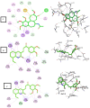
Fig. 12. 2D and 3D interaction diagrams of (A) colchicine, (B) compound 3b and (C) compound 3c into the colchicine binding site of tubulin enzyme.
Fig. 13
Fig. 13. 2D and 3D interaction diagrams of (A) compound 4c, (B) compound 5c into the colchicine binding site of tubulin enzyme and (C) aligned docking pose of colchicine (pink) and 4c into the CBS.
Fig. 14
Fig. 14. Radar chart showing six predicted physicochemical properties of the tested compound 3b, 3c, 4c, 5c, colchicine and CA-4.
Fig. 15
Fig. 15. Boiled egg model of the tested compounds 3b, 3c, 4c, 5c, colchicine and CA-4.
Fig. 16
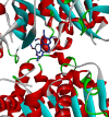
Fig. 16. Alignment between the conformation of the co-crystallized ligand (colchicine) colored in grey and the best-docked pose (colored in blue) into α–β tubulin enzyme with RMSD value equal to 0.17 Å.

Similar articles

Cited by

References

    1. Malabadi R. B. Sadiya M. Kolkar K. P. Mammadova S. S. Chalannavar R. K. Baijnath H. Int. J. Sci. Res. Arch. 2024;11:2502–2539. doi: 10.30574/ijsra.2024.11.1.0315. - DOI
    1. Łukasiewicz S. Czeczelewski M. Forma A. Baj J. Sitarz R. Stanisławek A. Cancers. 2021;13:1–30. doi: 10.3390/cancers13174287. - DOI - PMC - PubMed
    1. Sung H. Ferlay J. Siegel R. L. Laversanne M. Soerjomataram I. Jemal A. Bray F. Ca-Cancer J. Clin. 2021;71(3):209–249. doi: 10.3322/caac.21660. - DOI - PubMed
    1. Mukhtar E. Adhami V. M. Mukhtar H. Mol. Cancer Ther. 2014;13:275–284. doi: 10.1158/1535-7163.MCT-13-0791. - DOI - PMC - PubMed
    1. Lu Y. Chen J. Xiao M. Li W. Miller D. D. Pharm. Res. 2012;29:2943–2971. doi: 10.1007/s11095-012-0828-z.An. - DOI - PMC - PubMed

LinkOut - more resources