[Preprint]. 2024 Jul 4:2024.07.03.601941.
doi: 10.1101/2024.07.03.601941.

IL-13 decreases susceptibility to airway epithelial SARS-CoV-2 infection but increases disease severity in vivo

Affiliations

IL-13 decreases susceptibility to airway epithelial SARS-CoV-2 infection but increases disease severity in vivo

Shreya Ghimire et al. bioRxiv. .

Abstract

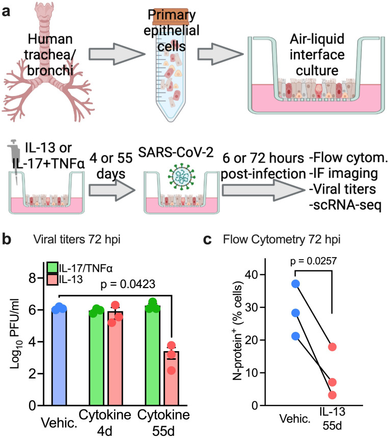
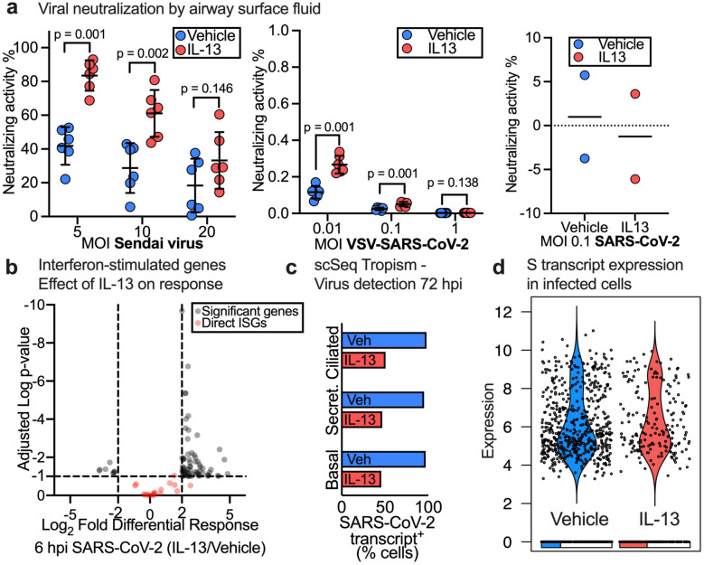
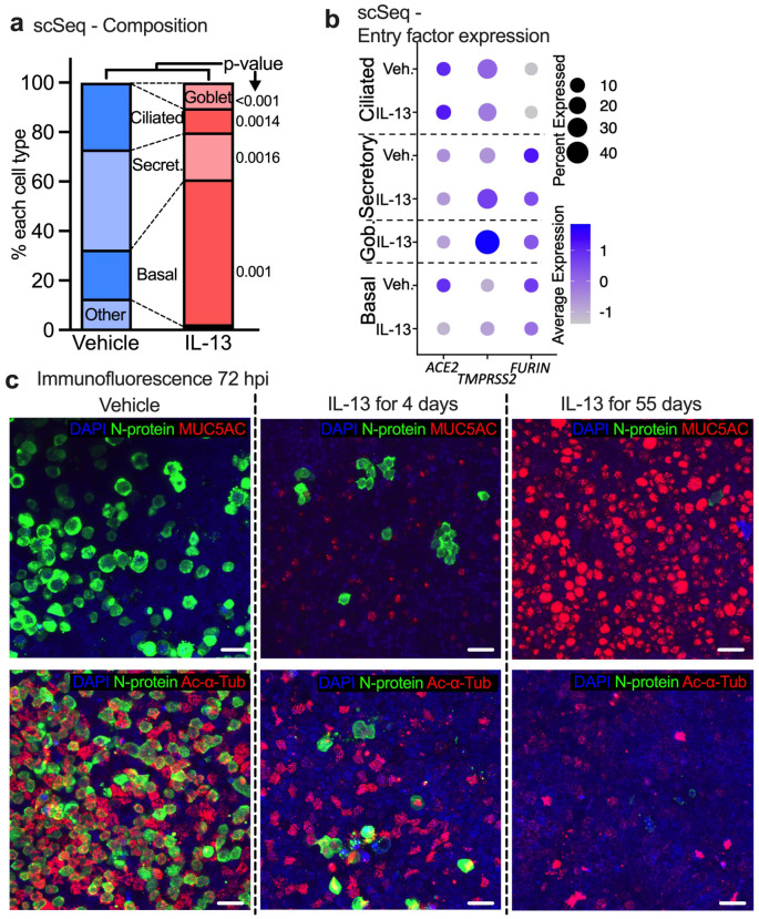
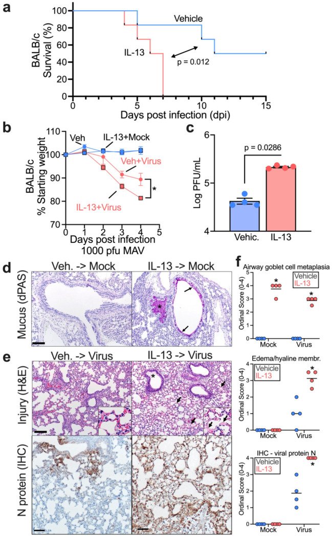
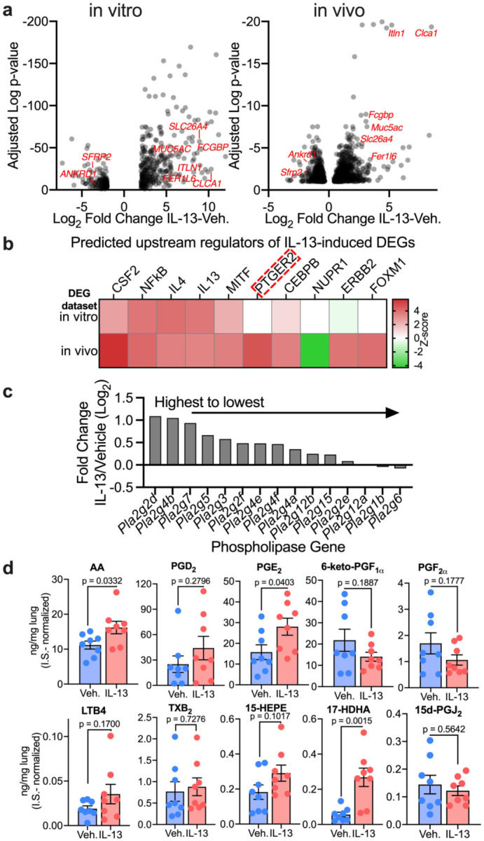
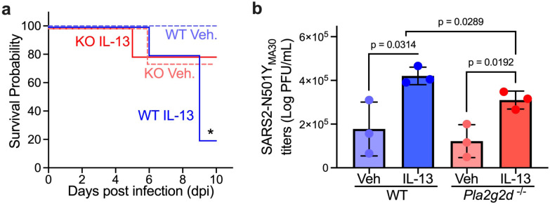
Treatments available to prevent progression of virus-induced lung diseases, including coronavirus disease 2019 (COVID-19) are of limited benefit once respiratory failure occurs. The efficacy of approved and emerging cytokine signaling-modulating antibodies is variable and is affected by disease course and patient-specific inflammation patterns. Therefore, understanding the role of inflammation on the viral infectious cycle is critical for effective use of cytokine-modulating agents. We investigated the role of the type 2 cytokine IL-13 on SARS-CoV-2 binding/entry, replication, and host response in primary HAE cells in vitro and in a model of mouse-adapted SARS-CoV-2 infection in vivo. IL-13 protected airway epithelial cells from SARS-CoV-2 infection in vitro by decreasing the abundance of ACE2-expressing ciliated cells rather than by neutralization in the airway surface liquid or by interferon-mediated antiviral effects. In contrast, IL-13 worsened disease severity in mice; the effects were mediated by eicosanoid signaling and were abolished in mice deficient in the phospholipase A2 enzyme PLA2G2D. We conclude that IL-13-induced inflammation differentially affects multiple steps of COVID-19 pathogenesis. IL-13-induced inflammation may be protective against initial SARS-CoV-2 airway epithelial infection; however, it enhances disease progression in vivo. Blockade of IL-13 and/or eicosanoid signaling may be protective against progression to severe respiratory virus-induced lung disease.

Keywords: COVID-19; IL-13; SARS-CoV-2; eicosanoids; prostaglandin; scRNA-seq.


Conflict of interest statement

Conflict of Interest: The authors have declared that no conflict of interest exists.

Figures

Figure 1.
Figure 1.. IL-13 induced inflammation protects human airway epithelia from SARS-CoV-2 infection in vitro.
(a) Experimental conditions/in-vitro model: IL-13 (20ng/mL) or IL-17 (20ng/mL) +TNFα (10ng/mL) were added to well-differentiated air-liquid interface (ALI) primary human airway epithelia (HAE) for 4 or 55 days prior to infection with SARS-CoV-2(MOI 0.1). Samples were analyzed at 6 or 72 hours post infection (hpi). (b) Viral titers/PFU at 72hpi. Data represent the mean ± SEM and p value for one-way ANOVA test. n =3 (c) Flow cytometry showing % of cells expressing N protein at 72hpi. n= epithelia from 3 human donors, p value shown for paired t-test. (a) was created with Biorender.com
Figure 2.
Figure 2.. IL-13 induced goblet cell metaplasia (GCM) does not affect viral neutralization at the airway surface or viral replication.
(a) Neutralization of Sendai virus (left), SARS-CoV-2 pseudo typed VSV (middle) and WT-SARS-CoV-2 (right) by airway surface liquid (ASL) collected from Vehicle and IL-13 treated (20ng/mL) HAE. n= epithelia from 6 or 2 humans, p-value shown for Student's unpaired t test and data represent mean ± SD. (b) Volcano plot for IL-13 (20ng/mL, 55 days) vs Vehicle pre-treated ciliated cells at 6 hpi with SARS-CoV-2. Dotted line represents cutoffs for significant genes (log2FC ± 2, adj p-value=0.1). Significant genes and ISGs directly affected by virus shown. (c) Effect of IL-13 treatment (20ng/mL, 55 days) on detection of SARS-CoV-2 transcript-positive cells in each major epithelial cell type. (d) Violin plot of S transcript in Vehicle vs IL-13 (20ng/mL, 55 days) treated HAE at 72 hpi with SARS-CoV-2. Each dot represents a SARS-CoV-2 transcript-positive cell and the bar at the bottom shows the proportion of cells with zero expression in each group.
Figure 3.
Figure 3.. IL-13 induced GCM protects HAE from SARS-CoV-2 in vitro by decreasing the abundance of receptor-expressing cells.
(a) Proportion of common cell types in vehicle- and IL-13-treated (20ng/mL, 55 days) samples (no virus exposure) determined by single cell RNA-seq data. n=3, p-value shown for unpaired t test. (b) Dot plot showing the effect of IL-13 treatment (20ng/mL, 55 days) on viral entry factor expression in uninfected HAE. Size of the dot represents % of the cell expressing the gene and color scale shows the average expression level. n = 3 donors (c) Representative image of immunofluorescence confocal microscopy for DAPI (nuclei, blue), N-protein (virus, green), and MUC5AC (goblet cells, red, top) or acetylated alpha tubulin (ciliated cells, red, bottom).
Figure 4.
Figure 4.. IL-13 enhances SARS2-N501YMA30-induced disease in vivo.
6-10 week-old BALB/c mice were intranasally infected by 1,000 PFU of SARS2-N501YMA30 after a 4-day pretreatment of 2.5 μg/day intranasal IL-13 or vehicle. (a) Survival rate and (b) weight were monitored daily. n = 4-6 mice for each group; mean ± SEM; for % starting weight of Veh + Virus vs IL-13+Virus, *p<0.05; p-values in weight change and survival curve are Student's unpaired t test and log-rank (Mantel-Cox) tests, respectively. (c) Quantification of virus loads at 5 dpi in homogenized mouse lungs by plaque assay using VeroE6 cells. n = lung tissue from 4 mice; p value: Student's unpaired t test. (d) Representative sections from uninfected mouse lungs treated with vehicle or IL-13treatment and stained with diastase-pretreated periodic acid-Schiff (dPAS). Note the increased dPAS+ staining of the epithelium (arrows) and within the airway (asterisk), (e) Representative sections from infected mouse lungs with vehicle or IL-13 treatment and stained with H&E (top) or immunostained for SARS-CoV-2 N protein. Note the edema (asterisk, top right image) and hyaline membranes (arrows, top right image). Note the expansive N protein staining in the bottom right versus bottom left representative images. Histopathology staining from 4 mice/group. (f) Ordinal histopathological scoring of airway goblet cell metaplasia (mucus-producing goblet cells) (top panel), edema/hyaline membrane (middle panel), and SARS-CoV-2 N protein expression (bottom panel) in lungs from mock or infected mice. mean ± SEM; *p<0.05; p value: Student's unpaired t test
Figure 5.
Figure 5.. IL-13 induces dysregulation of eicosanoids in vivo.
(a) RNA-seq analysis of IL-13 (20ng/mL)- or vehicle-treated primary human airway epithelial cells (21-day IL-13/Vehicle treatment, in vitro) and lungs of 6-10 week-old BALB/c mice (4-day, 2.5 μg/day intranasal IL-13/Vehicle treatment, in vivo). Volcano plots of differential gene expression analysis in vitro and in vivo of the effect of IL-13 compared to vehicle. n = epithelia from 8 humans (left); n = lung tissue from 4 mice (right). Selected genes relevant to goblet cell metaplasia are labeled in red. (b) Ingenuity Upstream Regulator Analysis of DEGs in IL-13- or vehicle-treated samples in vitro and in vivo. Predicted upstream regulators are sorted by absolute Z-score. PTGER2 is highlighted as the top predicted regulator activated in vivo but not in vitro. (c) List of top 15 eicosanoids biosynthesis-associated phospholipase genes ranked from highest to lowest magnitude of IL-13 response. (d) Concentrations of prostaglandins in lungs of 8 week-old BALB/c mice treated with IL-13 or vehicle measured by LC-MS/MS calculated according to standard curves and normalized to internal standards (0.1 ng/μL for each I.S.). n = lung sample from 8 mice; mean ± SEM; p value is shown from Student's unpaired t test.
Figure 6.
Figure 6.. IL-13 enhances SARS2-N501YMA30-induced disease in vivo via eicosanoid dysregulation.
8-9 month-old C57BL/6J wild-type (WT) and PLA2G2D-knock-out (KO) mice were pretreated with IL-13 and vehicle for 4 days and infected with 1,000 PFU SARS2-N501YMA30 on day 5. (a) Survival curve of infected wild type and PLA2G2D-knock-out mice. n = 4-5 mice for each group; mean ± SEM; *p<0.05; p value is shown from log-rank (Mantel-Cox) tests for WT Vehicle vs WT IL-13. (b) Viral loads in lungs of infected WT and KO mice on day 2 post infection were assessed by plaque assay. n = lung sample from 3 mice; mean ± SEM; p value is shown from Student's unpaired t test.

Similar articles

References

    1. Dryden-Peterson S, Kim A, Kim AY, Caniglia EC, Lennes IT, Patel R, et al. Nirmatrelvir Plus Ritonavir for Early COVID-19 in a Large U.S. Health System : A Population-Based Cohort Study. Ann Intern Med. 2023;176(1):77–84. - PMC - PubMed
    1. Matthay MA, and Luetkemeyer AF. IL-6 Receptor Antagonist Therapy for Patients Hospitalized for COVID-19: Who, When, and How? JAMA. 2021;326(6):483–5. - PubMed
    1. Zaid Y, Dore E, Dubuc I, Archambault AS, Flamand O, Laviolette M, et al. Chemokines and eicosanoids fuel the hyperinflammation within the lungs of patients with severe COVID-19. J Allergy Clin Immunol. 2021;148(2):368–80 e3. - PMC - PubMed
    1. Donlan AN, Sutherland TE, Marie C, Preissner S, Bradley BT, Carpenter RM, et al. IL-13 is a driver of COVID-19 severity. JCI Insight. 2021;6(15). - PMC - PubMed
    1. Donlan AN, Mallawaarachchi I, Sasson JM, Preissner R, Loomba JJ, and Petri WA. Dupilumab Use Is Associated With Protection From Coronavirus Disease 2019 Mortality: A Retrospective Analysis. Clin Infect Dis. 2023;76(1):148–51. - PMC - PubMed

Publication types