[Preprint]. 2024 Jul 4:2024.07.03.601953.
doi: 10.1101/2024.07.03.601953.

The Staphylococcus aureus small non-coding RNA IsrR regulates TCA cycle activity and virulence

Affiliations

The Staphylococcus aureus small non-coding RNA IsrR regulates TCA cycle activity and virulence

Gustavo Rios-Delgado et al. bioRxiv. .

Update in

Abstract

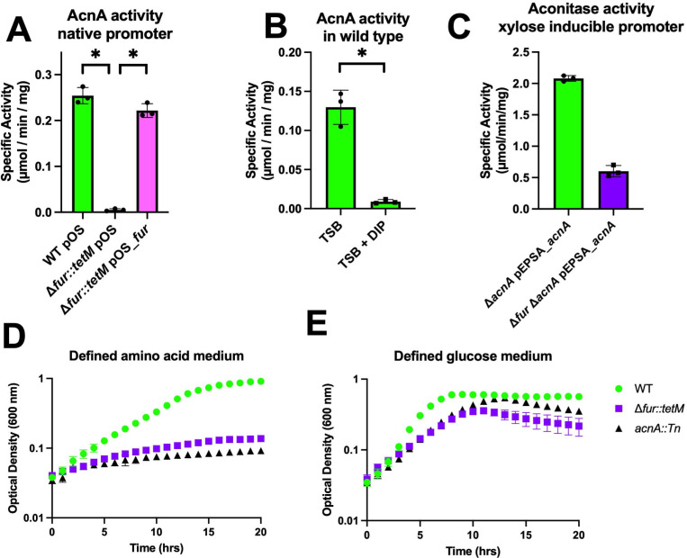
Staphylococcus aureus has evolved mechanisms to cope with low iron (Fe) availability in host tissues. S. aureus uses the ferric uptake transcriptional regulator (Fur) to sense titers of cytosolic Fe. Upon Fe depletion, apo-Fur relieves transcriptional repression of genes utilized for Fe uptake. We demonstrate that an S. aureus Δfur mutant has decreased expression of acnA, which codes for the Fe-dependent enzyme aconitase. Decreased acnA expression prevented the Δfur mutant from growing with amino acids as sole carbon and energy sources. Suppressor analysis determined that a mutation in isrR, which produces a regulatory RNA, permitted growth by decreasing isrR transcription. The decreased AcnA activity of the Δfur mutant was partially relieved by an ΔisrR mutation. Directed mutation of bases predicted to facilitate the interaction between the acnA transcript and IsrR, decreased the ability of IsrR to control acnA expression in vivo and IsrR bound to the acnA transcript in vitro. IsrR also bound to the transcripts coding the alternate TCA cycle proteins sdhC, mqo, citZ, and citM. Whole cell metal analyses suggest that IsrR promotes Fe uptake and increases intracellular Fe not ligated by macromolecules. Lastly, we determined that Fur and IsrR promote infection using murine skin and acute pneumonia models.

Keywords: Fur; IsrR; Staphylococcus aureus; Tsr25; iron.


Figures

Figure 1.
Figure 1.
A S. aureus Δfur mutant has decreased aconitase expression. Panel A. Aconitase activity was quantified in cell free lysates harvested from the wild type (WT) (JMB1100) and Δfur::tetM (JMB10842) strains carrying pOS-1-plgt or pOS-1-plgt_fur after culture in TSB-Cm medium. Panel B. Aconitase activity was quantified in cell free lysates from the WT after culture in TSB +/− 250 μM 2,2’ dipyridyl (DIP). Panel C. Aconitase activity was quantified in cell free lysates harvested from the acnA::Tn (JMB11803) and Δfur::tetM acnA::Tn (JMB11804) strains with pEPSA5_acnA after culture in TSB-Cm medium supplemented with 0.25% xylose. Panel D. Culture optical densities (A600) were monitored for the WT, Δfur::tetM, and acnA::Tn strains in a defined medium containing amino acids as the sole carbon and energy sources. Panel E. Culture optical densities (A600) of the WT, Δfur::tetM, and acnA::Tn strains were monitored in a liquid defined medium containing amino acids supplemented with 10 mM glucose. The data shown represent the average of biological triplicates with standard deviations shown. Error bars are shown for all data but in some cases (Panels D and E) are smaller than the symbols used. Student’s two-tailed t-tests were performed on the data and * represents a p-value of <0.05.
Figure 2.
Figure 2.
A null mutation in isrR suppresses the amino acid growth defect and increases aconitase activity of a Δfur mutant. Panel A. Culture optical densities (A600) of the wild type (WT) (JMB1100), Δfur::tetM (JMB10842), Δfur::tetM isrR* (JMB10495), and acnA::Tn (JMB11803) strains were monitored when cultured on defined medium containing amino acids as carbon and energy sources. Panel B. Aconitase activity was quantified in cell free lysates harvested from the WT, Δfur::tetM, and Δfur::tetM isrR* strains after culture in TSB medium. Panel C. Culture optical densities (A600) of the WT, acnA::Tn, Δfur::tetM, Δfur::tetM ΔisrR (JMB11293), and Δfur::tetM ΔisrR acnA::Tn (JMB11806) strains were monitored in liquid defined medium containing amino acids as carbon and energy sources. Panel D. Aconitase activity was quantified in cell free lysates harvested from the WT with pLL39 (JMB1886), SAUSA300_1456::Tn (1456::Tn) pLL39 (JMB11448), fur* 1456::Tn pLL39 (JMB11449), ΔisrR 1456::Tn pLL39 (JMB11395), fur* ΔisrR 1456::Tn pLL39 (JMB11392), and fur* ΔisrR 1456::Tn pLL39_isrR (JMB11393) strains after culture in TSB medium. E. Aconitase activity was quantified in cell free lysates harvested from the acnA::Tn, Δfur::tetM acnA::Tn (JMB11804), and Δfur::tetM ΔisrR acnA::Tn strains containing pEPSA5_acnA after culture in TSB-Cm medium supplemented with 0.25% xylose. The data shown represent the average of biological triplicates with standard deviations shown. Error bars are shown for all data but in some cases (Panels D and E) are smaller than the symbols used. Student’s two-tailed t-tests were performed on the data and * represents a p-value of <0.05.
Figure 3.
Figure 3.
The isrR* mutation suppresses the phenotypes of the Δfur mutant by decreasing isrR transcription. Panel A. Top, predicted consensus Fur box sequence as determined by RegPrecise, and bottom, consensus Staphylococcal Fur box, isrR distal Fur box, isrR proximal Fur box, and proximal Fur box in the isrR* strains with change highlighted and underlined. Panel B. Northern blot analysis of isrR transcripts using total RNA isolated from wild type (WT)(JMB1100) and ΔisrR (JMB11292) strains after culture in TSB media containing 0, 120, or 500 μM 2,2’ dipyridyl (DIP). Panel C. Northern blot analysis of isrR transcripts using total RNA isolated from WT, Δfur::tetM (JMB10842), and Δfur::tetM ΔisrR (JMB11293) strains after culture in TSB media with or without 120 μM DIP. Panel D. IsrR transcript abundance in the WT, Δfur::tetM, and Δfur::tetM isrR* strains after culture in liquid TSB media supplemented with or without 120 μM DIP. Transcript abundance was determined by quantitative PCR. Panel E. Quantification of transcripts corresponding to isrR after the Δfur::tetM ΔisrR strain carrying either pEPSA5_isrR or pEPSA5_isrR* were cultured in TSB medium containing 2% xylose and subsequently rifampicin was added (t=0) to inhibit transcription. Transcript abundances were normalized to t=0. Panel F. Relative fluorescence of the WT strain containing the pOS_pisrR_gfp or pOS_pisrR*_gfp transcriptional reporter after culture in TSB-Cm with or without 250 μM DIP. Panel G. Aconitase activity in cell free lysates from the WT strain carrying pEPSA5, pEPSA5_isrR, pEPSA5_as_isrR, pEPSA5_isrR*, and pEPSA5_trunk_isrR after culture in TSB-Cm with or without 2% xylose. Panels B and C contain representative Northern blots. The data displayed in panels D-G represent the average of biological triplicates with standard deviations shown. Student’s two-tailed t-tests were performed on the data and * represents a p-value of <0.05.
Figure 4.
Figure 4.
IsrR directly influences acnA translation. Panel A. IntaRNA predicted interaction between IsrR and acnA mRNA. Predicted interaction includes the acnA Shine Dalgarno and the second cytosine-rich region (CRR_2) of IsrR. The acnA mRNA AUG start codon is underlined. Panel B. Relative fluorescence of the wild type (WT) (JMB1100) and ΔisrR (JMB11292) strains containing the pOS_plgt_acnA_gfp translational reporter after culture in TSB-Cm media with or without 120 μM DIP. Panel C. Relative fluorescence of the WT, ΔisrR, Δfur::tetM (JMB10842), Δfur::tetM ΔisrR (JMB11293), and Δfur::tetM isrR* (JMB10495) strains containing the pOS_plgt_acnA_gfp translational reporter after culture in TSB-Cm medium. Panel D. Electrophoretic mobility shift assay (EMSA) using 20,000 cpm of radiolabeled IsrR and 0–500 μM of the acnA transcript. Panel E. EMSA using 20,000 cpm of radiolabeled IsrR and 0–500 μM of the lukH transcript. The data displayed in panels B and C represent the average of biological triplicates with standard deviations shown. Student’s two-tailed t-tests were performed on the data and * represents a p-value of <0.05. Pictures of representative EMSA assays (n=2) are displayed in panels D and E.
Figure 5.
Figure 5.
Interactions between nucleotides of the acnA Shine Dalgarno sequence and a cytosine rich region (CCR) of IsrR may influence IsrR-mediated acnA translational repression. Panel A. Partial sequences of the acnA_gfp and acnA1_gfp translational reporters. Black indicates acnA 5’ untranslated region (UTR), red indicates the acnA Shine Dalgarno sequence, blue indicates the first two codons of acnA, green indicates the start of the gfp sequence, and an asterisk above the nucleotide denotes that they are predicted to interact with IsrR (Figure 4A). Yellow nucleotides in the lower sequence indicate nucleotide substitutions in the acnA1_gfp translational reporter. Panel B. Relative fluorescence of the wild type (WT) (JMB1100), Δfur::tetM (JMB10842), ΔisrR (JMB11292), and Δfur::tetM ΔisrR (JMB11293) strains containing the acnA_gfp or acnA1_gfp translational reporters cultured in TSB-Cm medium. Panel C. Portions of IsrR and IsrR variant sequences (isrR_C1, isrR_C2, isrR_C1_C2) that are predicted to interact with acnA mRNA. Red indicates nucleotides involved in the predicted interaction that includes cytosine-rich region one (CRR_1), blue are the nucleotides in the predicted interaction with CRR_2. Underlined nucleotides indicate the IsrR C-rich regions. Yellow indicates the nucleotide substitutions on the isrR variants. Panel D. Relative fluorescence of the 1456::Tn pLL39 (JMB11448), fur* ΔisrR 1456::Tn pLL39 (JMB11392), fur* ΔisrR 1456::Tn pLL39_isrR (JMB11393), fur* ΔisrR 1456::Tn pLL39_isrR_C1 (JMB13983), fur* ΔisrR 1456::Tn pLL39_isrR_C2 (JMB13984), and fur* ΔisrR 1456::Tn pLL39_isrR_C1_C2 (JMB14314) containing the pOS_plgt_acnA_gfp translational reporter. Panel E. Aconitase activity in cell free lysates harvested from the strains in panel D after culture in TSB medium. The data displayed in panels B, D, and E represent the average of biological triplicates with standard deviations shown. Student’s two-tailed t-tests were performed on the data and * represents a p-value of <0.05.
Figure 6.
Figure 6.
IsrR interacts with TCA cycle mRNAs and affects expression. Panel A. Electrophoretic mobility shift assay (EMSA) using 20,000 cpm of radiolabeled IsrR and 0–500 nM of the sdhC transcript. Panel B. EMSA using 20,000 cpm of radiolabeled IsrR and 0–500 nM of the mqo transcript. Panel C. EMSA using 20,000 cpm of radiolabeled IsrR and 0–500 nM of the citZ or citM transcripts. Panel D. Activity of succinate dehydrogenase (Sdh) was quantified in cell free lysates generated from the wild type (WT) (JMB1100), Δfur::tetM (JMB10842), ΔisrR (JMB11292), and Δfur::tetM ΔisrR (JMB11293) strains after culture in TSB medium. Panel E. Activity of malate quinone oxidoreductase (Mqo) in cell free lysates generated from the WT, Δfur::tetM, ΔisrR, and Δfur::tetM ΔisrR strains. Pictures of representative EMSA assays (n=2) are displayed in panels A-C. The data displayed in panels D and E represent the average of biological triplicates with standard deviations shown. Student’s two-tailed t-tests were performed on the data and * represents a p-value of <0.05.
Figure 7.
Figure 7.
IsrR impacts iron homeostasis. Panel A. The wild type (WT)(JMB1100), Δfur::tetM (JMB10842), ΔisrR (JMB11292), and Δfur::tetM ΔisrR (JMB11293) strains were plated as top agar overlays on solid TSA, followed by spotting 2.5 μg streptonigrin. Panel B. The WT strain containing pEPSA5_isrR or pEPSA_as_isrR were plated as top agar overlays on solid TSA-Cm with or without 2% xylose, followed by spotting 2.5 μg streptonigrin. For Panels A and B, the zones of clearance resulting from streptonigrin growth inhibition was quantified. Panels C, D, and E. The ratio of 56Fe and 24Mg abundances were quantified in whole cells using ICP-MS after culture in Chelex treated TSB (Panel C), TSB (Panel D), or TSB supplemented with 50 μM Fe (Panel E). The ratio of 56Fe / 24Mg is displayed for WT, Δfur::tetM, ΔisrR, and Δfur::tetM ΔisrR strains. Panel F. Siderophore production from spent culture supernatants from the WT, Δfur::tetM, ΔisrR, and Δfur::tetM ΔisrR strains was quantified. The data displayed represent the average of biological triplicates with standard deviations shown. Student’s two-tailed t-tests were performed on the data and * represents a p-value of <0.05.
Figure 8.
Figure 8.
IsrR and Fur are important for tissue damage and colonization during infection. Panels A and B. The wild type (WT)(JMB1100), Δfur::tetM (JMB10842), ΔisrR (JMB11292), and Δfur::tetM ΔisrR (JMB11293) strains were tested in a model of acute pneumonia infection. Data represent bacterial counts 24 hours after intranasal infection in bronchoalveolar lavage fluid (Panel A) and lung tissue (Panel B). Panels C and D, the wild type (WT), Δfur::tetM, ΔisrR, and Δfur::tetM ΔisrR strains were injected subcutaneously into C57BL/6J mice and total lesion size (Panel C) and necrosis size (Panel D) were monitored over time. Panel E. Local bacterial titers were quantified four days post infection. Data is a representative experiment with n=10. For A and B, each symbol represents the mean. For C and D, each dot is an individual animal, and the bar or line represents the mean. Error bars represent the SEM and may be smaller than symbols. * and ** indicates p<0.05 and p<0.01, respectively, compared to WT for Δfur::tetM and Δfur::tetM ΔisrR by Mann-Whitney test.
Figure 9.
Figure 9.
Model for IsrR-dependent regulation of TCA cycle expression. Growth in iron ion replete conditions results in metalation of Fur and transcriptional repression of isrR, which produces a non-coding small regulatory RNA. Upon iron limitation, Fur is demetallated, and isrR is expressed. IsrR forms complexes with the acnA, sdh, citM, and citZ mRNA transcripts resulting in decreased expression and decreased carbon flux through the TCA cycle. Decreased expression of TCA cycle enzymes results in an inability to grow using amino acids for carbon and energy. Figure was created using Biorender.

Similar articles

References

    1. Tong S.Y., Davis J.S., Eichenberger E., Holland T.L. and Fowler V.G. Jr. (2015) Staphylococcus aureus infections: epidemiology, pathophysiology, clinical manifestations, and management. Clin Microbiol Rev, 28, 603–661. - PMC - PubMed
    1. De Oliveira D.M.P., Forde B.M., Kidd T.J., Harris P.N.A., Schembri M.A., Beatson S.A., Paterson D.L. and Walker M.J. (2020) Antimicrobial Resistance in ESKAPE Pathogens. Clin Microbiol Rev, 33. - PMC - PubMed
    1. Mashruwala A.A., Pang Y.Y., Rosario-Cruz Z., Chahal H.K., Benson M.A., Mike L.A., Skaar E.P., Torres V.J., Nauseef W.M. and Boyd J.M. (2015) Nfu facilitates the maturation of iron-sulfur proteins and participates in virulence in Staphylococcus aureus. Mol Microbiol, 95, 383–409. - PMC - PubMed
    1. Skaar E.P., Humayun M., Bae T., DeBord K.L. and Schneewind O. (2004) Iron-source preference of Staphylococcus aureus infections. Science, 305, 1626–1628. - PubMed
    1. Torres V.J., Attia A.S., Mason W.J., Hood M.I., Corbin B.D., Beasley F.C., Anderson K.L., Stauff D.L., McDonald W.H., Zimmerman L.J. et al. (2010) Staphylococcus aureus Fur regulates the expression of virulence factors that contribute to the pathogenesis of pneumonia. Infect Immun, 78, 1618–1628. - PMC - PubMed

Publication types

LinkOut - more resources